Stepanka Homola, Sait Sendag, Davut Koca, Axel Wehrend
{"title":"Methods for Evaluating Testicular Function in Domestic Cats.","authors":"Stepanka Homola, Sait Sendag, Davut Koca, Axel Wehrend","doi":"10.1111/rda.70042","DOIUrl":null,"url":null,"abstract":"<p><p>This study aimed to describe the physiological sonomorphology of the cat testicle and to verify the findings by histological and endocrinological analyses. Furthermore, two methods of testicle measurement and volume calculation were compared. For the study, a clinical examination of the testicles was carried out in 39 cats. The testicles were measured with the Podany testimeter and using a sonographic examination. This was followed by a castration and histological examination of the testicle. The testosterone level was measured from a blood sample. The cat testicles showed a characteristic sonomorphology. The parenchyma was homogeneous, with a distinct echogenic mediastinum. The histological examination verified that there was spermatogenesis activity in testicular tissue. It was found that the testosterone concentrations varied greatly between the animals (0.1-4.40 ng/mL) although spermatogenesis was detected in all cats. Significant correlations were detected between right and left testicular volumes of the cats according to the measured values of sonography and Podany testimeter (p < 0.001). In addition, there was a significant and positive correlation between testosterone and testicular volume. Testosterone levels increase with increasing testicular volume according to the sonographic method (p < 0.05). With the help of the sonographic measurement, significantly higher testicular volumes are calculated than were compared with the measurement method with the Podany testimeter (p < 0.05). The present study is the first to provide detailed information and reliable data for the evaluation of the testicle size and volume in male cats as well as for sonomorphology, which can be used as comparison values for the andrological examination of this animal species.</p>","PeriodicalId":21035,"journal":{"name":"Reproduction in Domestic Animals","volume":"60 3","pages":"e70042"},"PeriodicalIF":1.7000,"publicationDate":"2025-03-01","publicationTypes":"Journal Article","fieldsOfStudy":null,"isOpenAccess":false,"openAccessPdf":"https://www.ncbi.nlm.nih.gov/pmc/articles/PMC11889521/pdf/","citationCount":"0","resultStr":null,"platform":"Semanticscholar","paperid":null,"PeriodicalName":"Reproduction in Domestic Animals","FirstCategoryId":"97","ListUrlMain":"https://doi.org/10.1111/rda.70042","RegionNum":3,"RegionCategory":"农林科学","ArticlePicture":[],"TitleCN":null,"AbstractTextCN":null,"PMCID":null,"EPubDate":"","PubModel":"","JCR":"Q2","JCRName":"AGRICULTURE, DAIRY & ANIMAL SCIENCE","Score":null,"Total":0}
引用次数: 0
Abstract
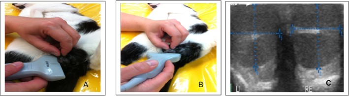
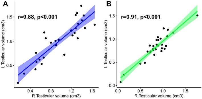
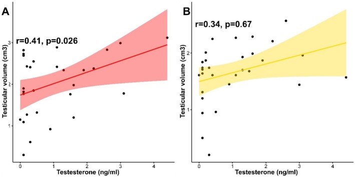
This study aimed to describe the physiological sonomorphology of the cat testicle and to verify the findings by histological and endocrinological analyses. Furthermore, two methods of testicle measurement and volume calculation were compared. For the study, a clinical examination of the testicles was carried out in 39 cats. The testicles were measured with the Podany testimeter and using a sonographic examination. This was followed by a castration and histological examination of the testicle. The testosterone level was measured from a blood sample. The cat testicles showed a characteristic sonomorphology. The parenchyma was homogeneous, with a distinct echogenic mediastinum. The histological examination verified that there was spermatogenesis activity in testicular tissue. It was found that the testosterone concentrations varied greatly between the animals (0.1-4.40 ng/mL) although spermatogenesis was detected in all cats. Significant correlations were detected between right and left testicular volumes of the cats according to the measured values of sonography and Podany testimeter (p < 0.001). In addition, there was a significant and positive correlation between testosterone and testicular volume. Testosterone levels increase with increasing testicular volume according to the sonographic method (p < 0.05). With the help of the sonographic measurement, significantly higher testicular volumes are calculated than were compared with the measurement method with the Podany testimeter (p < 0.05). The present study is the first to provide detailed information and reliable data for the evaluation of the testicle size and volume in male cats as well as for sonomorphology, which can be used as comparison values for the andrological examination of this animal species.
期刊介绍:
The journal offers comprehensive information concerning physiology, pathology, and biotechnology of reproduction. Topical results are currently published in original papers, reviews, and short communications with particular attention to investigations on practicable techniques.
Carefully selected reports, e. g. on embryo transfer and associated biotechnologies, gene transfer, and spermatology provide a link between basic research and clinical application. The journal applies to breeders, veterinarians, and biologists, and is also of interest in human medicine. Interdisciplinary cooperation is documented in the proceedings of the joint annual meetings.
Fields of interest: Animal reproduction and biotechnology with special regard to investigations on applied and clinical research.
 求助内容:
求助内容: 应助结果提醒方式:


