Dougal Ferguson, Niels Kroeger-Lui, Domenic Dreisbach, Claire A. Hart, Diego F. Sanchez, Pedro Oliveira, Mick Brown, Noel Clarke, Ashwin Sachdeva and Peter Gardner
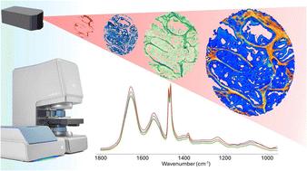
{"title":"Full fingerprint hyperspectral imaging of prostate cancer tissue microarrays within clinical timeframes using quantum cascade laser microscopy†","authors":"Dougal Ferguson, Niels Kroeger-Lui, Domenic Dreisbach, Claire A. Hart, Diego F. Sanchez, Pedro Oliveira, Mick Brown, Noel Clarke, Ashwin Sachdeva and Peter Gardner","doi":"10.1039/D5AN00046G","DOIUrl":null,"url":null,"abstract":"<p >One of the major limitations for clinical applications of infrared spectroscopic imaging modalities is the acquisition time required to obtain reasonable images of tissues with high spatial resolution and good signal-to-noise ratio (SNR). The time to acquire a reasonable signal to noise spectroscopic scan of a standard microscope slide region of tissue can take many hours. As a trade-off, systems can allow for discrete wavenumber acquisitions, sacrificing potentially vital chemical bands in order to reach specific acquisition targets. Recent instrumentation developments now allow for the full fingerprint imaging of entire microscope slides in under 30 minutes, enabling rapid, high quality spectroscopic imaging of tissues within clinical timeframes without sacrificing frequency bands. Here we compare the data from a novel QCL microscope to an FTIR microscope covering multiple aspects of spectroscopic imaging of a large, clinically relevant, prostate cancer tissue cohort (<em>N</em> = 1281). Comparisons of hyperspectral data acquisition quality in both achieved signal to noise and image contrast alongside the capacity for unsupervised and supervised modelling of tissue constituents are reported. We conclude that it is now possible to collect full fingerprint spectra and derive clinically relevant data in a timeframe suitable for translation into the pathology laboratory without the need to resort to discrete frequency imaging with subsequent loss of information.</p>","PeriodicalId":63,"journal":{"name":"Analyst","volume":" 9","pages":" 1741-1753"},"PeriodicalIF":3.3000,"publicationDate":"2025-03-10","publicationTypes":"Journal Article","fieldsOfStudy":null,"isOpenAccess":false,"openAccessPdf":"https://pubs.rsc.org/en/content/articlepdf/2025/an/d5an00046g?page=search","citationCount":"0","resultStr":null,"platform":"Semanticscholar","paperid":null,"PeriodicalName":"Analyst","FirstCategoryId":"92","ListUrlMain":"https://pubs.rsc.org/en/content/articlelanding/2025/an/d5an00046g","RegionNum":3,"RegionCategory":"化学","ArticlePicture":[],"TitleCN":null,"AbstractTextCN":null,"PMCID":null,"EPubDate":"","PubModel":"","JCR":"Q2","JCRName":"CHEMISTRY, ANALYTICAL","Score":null,"Total":0}
引用次数: 0
Abstract
One of the major limitations for clinical applications of infrared spectroscopic imaging modalities is the acquisition time required to obtain reasonable images of tissues with high spatial resolution and good signal-to-noise ratio (SNR). The time to acquire a reasonable signal to noise spectroscopic scan of a standard microscope slide region of tissue can take many hours. As a trade-off, systems can allow for discrete wavenumber acquisitions, sacrificing potentially vital chemical bands in order to reach specific acquisition targets. Recent instrumentation developments now allow for the full fingerprint imaging of entire microscope slides in under 30 minutes, enabling rapid, high quality spectroscopic imaging of tissues within clinical timeframes without sacrificing frequency bands. Here we compare the data from a novel QCL microscope to an FTIR microscope covering multiple aspects of spectroscopic imaging of a large, clinically relevant, prostate cancer tissue cohort (N = 1281). Comparisons of hyperspectral data acquisition quality in both achieved signal to noise and image contrast alongside the capacity for unsupervised and supervised modelling of tissue constituents are reported. We conclude that it is now possible to collect full fingerprint spectra and derive clinically relevant data in a timeframe suitable for translation into the pathology laboratory without the need to resort to discrete frequency imaging with subsequent loss of information.
 求助内容:
求助内容: 应助结果提醒方式:


