Paula Terra Bandeira, Camila Rodrigues Chaves, Pedro Henrique Monteiro Torres, Wanderley de Souza
{"title":"Immunolocalization and 3D modeling of three unique proteins belonging to the costa of Tritrichomonas foetus.","authors":"Paula Terra Bandeira, Camila Rodrigues Chaves, Pedro Henrique Monteiro Torres, Wanderley de Souza","doi":"10.1007/s00436-025-08466-4","DOIUrl":null,"url":null,"abstract":"<p><p>Nowadays, even in light of all the massive advances in cell biology, we still find some cellular structures that are not entirely understood. Among those, we highlight the costa, a structure from the mastigont system existent only in some members of the orders Trichomonadida and Tritrichomonadida, including the pathogens of venereal diseases in humans and cattle, Trichomonas vaginalis (T. vaginalis) and Tritrichomonas foetus (T. foetus), respectively. The costa is a prominent striated fiber and, although part of the cytoskeleton, differs from its classical components, and its molecular composition is still not fully characterized. Using proteomics of T. foetus's costa fraction, we previously identified hypothetic proteins, and among these, the protein ARM19800.1 positively localized in the costa and named costain-1. In this study, two other protein candidates were analyzed. To achieve the specific localization of 11810 and 32137 proteins in T. foetus's cells, it was used expansion microscopy and immunocytochemistry. The immunofluorescence revealed the presence of both proteins throughout the whole costa but with different intensities. Immunocytochemistry using negative staining, LR-White, and Epon embedding revealed further analyses of the protein's localization. All techniques confirmed the distinct and distributed localization of both proteins: costain-2 (11810) and costain-3 (32137). Also, AlfaFold3 was used to generate 3D models of the three identified proteins, showing a major prevalence of α-helical spans. Nonetheless, the identification and further characterization of these unique proteins can help understand their functional role in the assembled costa and, therefore, better understand the organization and function of this structure in these organisms.</p>","PeriodicalId":19968,"journal":{"name":"Parasitology Research","volume":"124 3","pages":"30"},"PeriodicalIF":2.0000,"publicationDate":"2025-03-07","publicationTypes":"Journal Article","fieldsOfStudy":null,"isOpenAccess":false,"openAccessPdf":"https://www.ncbi.nlm.nih.gov/pmc/articles/PMC11889022/pdf/","citationCount":"0","resultStr":null,"platform":"Semanticscholar","paperid":null,"PeriodicalName":"Parasitology Research","FirstCategoryId":"3","ListUrlMain":"https://doi.org/10.1007/s00436-025-08466-4","RegionNum":3,"RegionCategory":"医学","ArticlePicture":[],"TitleCN":null,"AbstractTextCN":null,"PMCID":null,"EPubDate":"","PubModel":"","JCR":"Q2","JCRName":"PARASITOLOGY","Score":null,"Total":0}
引用次数: 0
Abstract
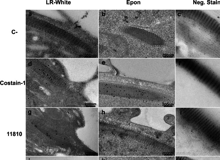
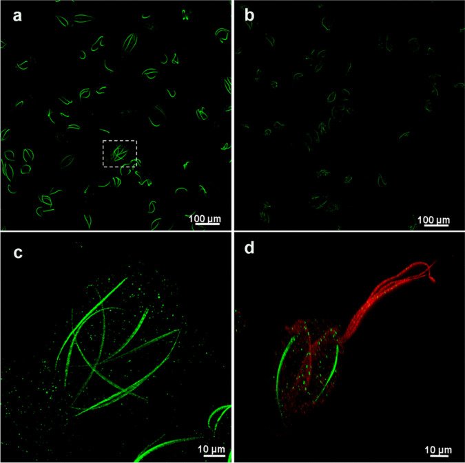
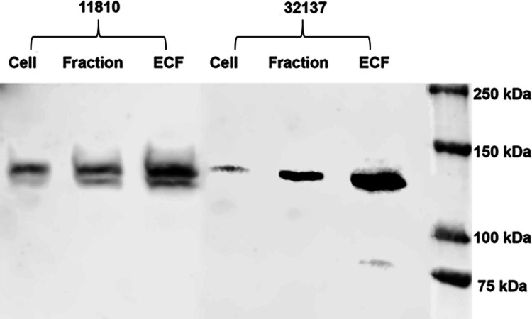
Nowadays, even in light of all the massive advances in cell biology, we still find some cellular structures that are not entirely understood. Among those, we highlight the costa, a structure from the mastigont system existent only in some members of the orders Trichomonadida and Tritrichomonadida, including the pathogens of venereal diseases in humans and cattle, Trichomonas vaginalis (T. vaginalis) and Tritrichomonas foetus (T. foetus), respectively. The costa is a prominent striated fiber and, although part of the cytoskeleton, differs from its classical components, and its molecular composition is still not fully characterized. Using proteomics of T. foetus's costa fraction, we previously identified hypothetic proteins, and among these, the protein ARM19800.1 positively localized in the costa and named costain-1. In this study, two other protein candidates were analyzed. To achieve the specific localization of 11810 and 32137 proteins in T. foetus's cells, it was used expansion microscopy and immunocytochemistry. The immunofluorescence revealed the presence of both proteins throughout the whole costa but with different intensities. Immunocytochemistry using negative staining, LR-White, and Epon embedding revealed further analyses of the protein's localization. All techniques confirmed the distinct and distributed localization of both proteins: costain-2 (11810) and costain-3 (32137). Also, AlfaFold3 was used to generate 3D models of the three identified proteins, showing a major prevalence of α-helical spans. Nonetheless, the identification and further characterization of these unique proteins can help understand their functional role in the assembled costa and, therefore, better understand the organization and function of this structure in these organisms.
期刊介绍:
The journal Parasitology Research covers the latest developments in parasitology across a variety of disciplines, including biology, medicine and veterinary medicine. Among many topics discussed are chemotherapy and control of parasitic disease, and the relationship of host and parasite.
Other coverage includes: Protozoology, Helminthology, Entomology; Morphology (incl. Pathomorphology, Ultrastructure); Biochemistry, Physiology including Pathophysiology;
Parasite-Host-Relationships including Immunology and Host Specificity; life history, ecology and epidemiology; and Diagnosis, Chemotherapy and Control of Parasitic Diseases.
 求助内容:
求助内容: 应助结果提醒方式:


