Seyed Mohsen Rafizadeh, Amir Mousavi, Mohammad Taher Rajabi, Amirhossein Aghajani, Zohreh Nozarian, Amin Zand
{"title":"Orbital and medial rectus muscle involvement as initial presentations of hydatid disease.","authors":"Seyed Mohsen Rafizadeh, Amir Mousavi, Mohammad Taher Rajabi, Amirhossein Aghajani, Zohreh Nozarian, Amin Zand","doi":"10.1186/s12348-025-00476-8","DOIUrl":null,"url":null,"abstract":"<p><strong>Purpose: </strong>To report a rare case of orbital hydatid cyst involving the medial rectus muscle, which presented as progressive proptosis, with subsequent detection of liver involvement after further investigations.</p><p><strong>Case presentation: </strong>We present the case of a 12-year-old boy from a rural area with exposure to wildlife dogs. The patient had a two-month history of gradually progressive proptosis in the right eye, accompanied by periorbital swelling and limited medial ocular motility. Orbital magnetic resonance imaging (MRI) revealed a large mass within the medial rectus muscle, which showed peripheral enhancement with no central enhancement, consistent with a cystic lesion based on its imaging characteristics. The patient underwent orbitotomy, during which the lesion was aspirated, and its walls were resected. Pathological examination confirmed a structure of a hydatid cyst. Given the suggestive signs of a hydatid cyst as part of a systemic echinococcal infection, further investigations, including liver sonography, revealed a similar cystic lesion in the hepatic lobe. With the diagnosis of an orbital hydatid cyst and suspected echinococcal infection, the patient was treated with oral Albendazole for one month. His symptoms, including periorbital swelling, improved, and no recurrence was observed at a six-month follow-up.</p><p><strong>Conclusions: </strong>Orbital hydatid cysts may present as inflammatory proptosis and should be considered in populations from endemic areas of human echinococcosis. Early diagnosis using orbital MRI, systemic investigations such as liver sonography, timely surgery for definitive diagnosis and treatment, and appropriate adjuvant antiparasitic medication are crucial for effective management.</p>","PeriodicalId":16600,"journal":{"name":"Journal of Ophthalmic Inflammation and Infection","volume":"15 1","pages":"21"},"PeriodicalIF":2.3000,"publicationDate":"2025-03-07","publicationTypes":"Journal Article","fieldsOfStudy":null,"isOpenAccess":false,"openAccessPdf":"https://www.ncbi.nlm.nih.gov/pmc/articles/PMC11889293/pdf/","citationCount":"0","resultStr":null,"platform":"Semanticscholar","paperid":null,"PeriodicalName":"Journal of Ophthalmic Inflammation and Infection","FirstCategoryId":"1085","ListUrlMain":"https://doi.org/10.1186/s12348-025-00476-8","RegionNum":0,"RegionCategory":null,"ArticlePicture":[],"TitleCN":null,"AbstractTextCN":null,"PMCID":null,"EPubDate":"","PubModel":"","JCR":"Q1","JCRName":"OPHTHALMOLOGY","Score":null,"Total":0}
引用次数: 0
Abstract
Purpose: To report a rare case of orbital hydatid cyst involving the medial rectus muscle, which presented as progressive proptosis, with subsequent detection of liver involvement after further investigations.
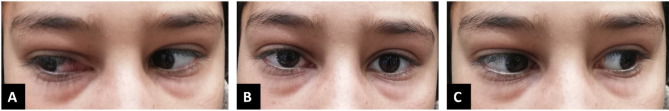
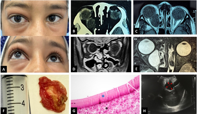
Case presentation: We present the case of a 12-year-old boy from a rural area with exposure to wildlife dogs. The patient had a two-month history of gradually progressive proptosis in the right eye, accompanied by periorbital swelling and limited medial ocular motility. Orbital magnetic resonance imaging (MRI) revealed a large mass within the medial rectus muscle, which showed peripheral enhancement with no central enhancement, consistent with a cystic lesion based on its imaging characteristics. The patient underwent orbitotomy, during which the lesion was aspirated, and its walls were resected. Pathological examination confirmed a structure of a hydatid cyst. Given the suggestive signs of a hydatid cyst as part of a systemic echinococcal infection, further investigations, including liver sonography, revealed a similar cystic lesion in the hepatic lobe. With the diagnosis of an orbital hydatid cyst and suspected echinococcal infection, the patient was treated with oral Albendazole for one month. His symptoms, including periorbital swelling, improved, and no recurrence was observed at a six-month follow-up.
Conclusions: Orbital hydatid cysts may present as inflammatory proptosis and should be considered in populations from endemic areas of human echinococcosis. Early diagnosis using orbital MRI, systemic investigations such as liver sonography, timely surgery for definitive diagnosis and treatment, and appropriate adjuvant antiparasitic medication are crucial for effective management.
 求助内容:
求助内容: 应助结果提醒方式:


