Pablo Naval-Baudin, Karen Pérez-Alfonso, Albert Castillo-Pinar, Ignacio Martínez-Zalacaín, Pablo Arroyo-Pereiro, Lucía Romero-Pinel, Nahum Calvo, Antonio Martinez-Yélamos, Mónica Cos, Sergio Martínez-Yélamos, Albert Pons-Escoda, Carles Majós
{"title":"Post-contrast Susceptibility Weighted Imaging in Multiple Sclerosis MRI Improves the Detection of Enhancing Lesions.","authors":"Pablo Naval-Baudin, Karen Pérez-Alfonso, Albert Castillo-Pinar, Ignacio Martínez-Zalacaín, Pablo Arroyo-Pereiro, Lucía Romero-Pinel, Nahum Calvo, Antonio Martinez-Yélamos, Mónica Cos, Sergio Martínez-Yélamos, Albert Pons-Escoda, Carles Majós","doi":"10.1007/s00062-025-01508-5","DOIUrl":null,"url":null,"abstract":"<p><strong>Objectives: </strong>MRI is essential for monitoring multiple sclerosis (MS). Contrast-enhanced T1-weighted imaging (T1WI+C) detects active inflammatory lesions indicating blood-brain barrier breakdown and is relevant for disease monitoring and treatment optimization. Susceptibility-weighted imaging (SWI) may be included in the imaging protocol for detecting MS-specific features, such as the presence of central veins or paramagnetic rim lesions. However, post-contrast SWI (SWI+C) has an inherent \"T1 shine-through effect\" that enables the visualization of contrast-enhancing lesions. This study evaluates whether SWI+C in addition to standard T1WI+C improves the detection of enhancing lesions in patients with MS.</p><p><strong>Materials and methods: </strong>The images of 310 patients with MS who underwent a standardized MRI protocol including T1WI+C and SWI+C using a 3T scanner were retrospectively reviewed. A neuroradiologist and radiology resident independently evaluated the images obtained on T1WI+C alone and T1WI+C plus SWI+C. The efficacy of T1WI+C alone was compared with that of T1WI+C plus SWI+C for detecting active enhancing MS lesions.</p><p><strong>Results: </strong>The neuroradiologist detected 117 lesions on T1WI+C and 123 lesions on T1WI+C plus SWI+C. The resident detected 108 lesions on T1WI+C and 121 lesions on T1WI+C plus SWI+C. The interobserver agreement improved from 0.981 to 1.00 with the addition of SWI+C.</p><p><strong>Conclusion: </strong>Adding SWI+C to standard T1WI+C consistently enhances the detection of active enhancing inflammatory MS lesions and the interobserver agreement. If standardized, this combined approach may allow for earlier detection of disease activity and improve monitoring of MS progression, potentially leading to optimized treatment decisions and improved patient outcomes.</p>","PeriodicalId":10391,"journal":{"name":"Clinical Neuroradiology","volume":" ","pages":"533-539"},"PeriodicalIF":2.6000,"publicationDate":"2025-09-01","publicationTypes":"Journal Article","fieldsOfStudy":null,"isOpenAccess":false,"openAccessPdf":"https://www.ncbi.nlm.nih.gov/pmc/articles/PMC12454511/pdf/","citationCount":"0","resultStr":null,"platform":"Semanticscholar","paperid":null,"PeriodicalName":"Clinical Neuroradiology","FirstCategoryId":"3","ListUrlMain":"https://doi.org/10.1007/s00062-025-01508-5","RegionNum":3,"RegionCategory":"医学","ArticlePicture":[],"TitleCN":null,"AbstractTextCN":null,"PMCID":null,"EPubDate":"2025/3/7 0:00:00","PubModel":"Epub","JCR":"Q2","JCRName":"Medicine","Score":null,"Total":0}
引用次数: 0
Abstract
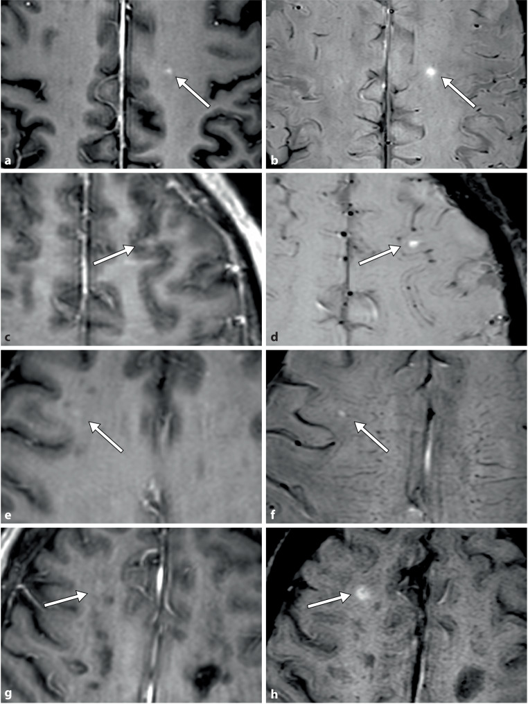
Objectives: MRI is essential for monitoring multiple sclerosis (MS). Contrast-enhanced T1-weighted imaging (T1WI+C) detects active inflammatory lesions indicating blood-brain barrier breakdown and is relevant for disease monitoring and treatment optimization. Susceptibility-weighted imaging (SWI) may be included in the imaging protocol for detecting MS-specific features, such as the presence of central veins or paramagnetic rim lesions. However, post-contrast SWI (SWI+C) has an inherent "T1 shine-through effect" that enables the visualization of contrast-enhancing lesions. This study evaluates whether SWI+C in addition to standard T1WI+C improves the detection of enhancing lesions in patients with MS.
Materials and methods: The images of 310 patients with MS who underwent a standardized MRI protocol including T1WI+C and SWI+C using a 3T scanner were retrospectively reviewed. A neuroradiologist and radiology resident independently evaluated the images obtained on T1WI+C alone and T1WI+C plus SWI+C. The efficacy of T1WI+C alone was compared with that of T1WI+C plus SWI+C for detecting active enhancing MS lesions.
Results: The neuroradiologist detected 117 lesions on T1WI+C and 123 lesions on T1WI+C plus SWI+C. The resident detected 108 lesions on T1WI+C and 121 lesions on T1WI+C plus SWI+C. The interobserver agreement improved from 0.981 to 1.00 with the addition of SWI+C.
Conclusion: Adding SWI+C to standard T1WI+C consistently enhances the detection of active enhancing inflammatory MS lesions and the interobserver agreement. If standardized, this combined approach may allow for earlier detection of disease activity and improve monitoring of MS progression, potentially leading to optimized treatment decisions and improved patient outcomes.
期刊介绍:
Clinical Neuroradiology provides current information, original contributions, and reviews in the field of neuroradiology. An interdisciplinary approach is accomplished by diagnostic and therapeutic contributions related to associated subjects.
The international coverage and relevance of the journal is underlined by its being the official journal of the German, Swiss, and Austrian Societies of Neuroradiology.
 求助内容:
求助内容: 应助结果提醒方式:


