Evaluation of CT and MRI Radiomics for an Early Assessment of Diffuse Axonal Injury in Patients with Traumatic Brain Injury Compared to Conventional Radiological Diagnosis.
Anna-Katharina Meißner, Robin Gutsche, Lenhard Pennig, Christian Nelles, Enrico Budzejko, Christina Hamisch, Martin Kocher, Marc Schlamann, Roland Goldbrunner, Stefan Grau, Philipp Lohmann
{"title":"Evaluation of CT and MRI Radiomics for an Early Assessment of Diffuse Axonal Injury in Patients with Traumatic Brain Injury Compared to Conventional Radiological Diagnosis.","authors":"Anna-Katharina Meißner, Robin Gutsche, Lenhard Pennig, Christian Nelles, Enrico Budzejko, Christina Hamisch, Martin Kocher, Marc Schlamann, Roland Goldbrunner, Stefan Grau, Philipp Lohmann","doi":"10.1007/s00062-025-01507-6","DOIUrl":null,"url":null,"abstract":"<p><strong>Background: </strong>De- and acceleration traumata can cause diffuse axonal injury (DAI) in patients with traumatic brain injury (TBI). The diagnosis of DAI on CT is challenging due to the lack of structural abnormalities. Radiomics, a method from the field of artificial intelligence (AI) offers the opportunity to extract additional information from imaging data. The purpose of this work was the evaluation of the feasibility of radiomics for an improved diagnosis of DAI in comparison to conventional radiological image assessment.</p><p><strong>Methods: </strong>CT and MR imaging was performed in 42 patients suspicious of DAI due to the clinical state, and two control groups (n = 44;42). DAI was diagnosed by experienced neuroradiologists. Radiomics features were extracted using a standardized MRI-based atlas of the predilection areas for DAI. Different MRI and CT based models were trained and validated by five-fold cross validation. Diagnostic performance was compared to the reading of two experienced radiologists and further validated in an external test dataset.</p><p><strong>Results: </strong>The MRI and CT models showed significant differences in radiomics features between patients with DAI and controls. The developed MRI based random forest classifier yielded an accuracy of 80-90%. The best performing CT model yielded an accuracy of 88% in the training data and 70% in the external test data. The results were comparable to conventional image analysis which achieved an accuracy of 70-81% for CT-based diagnosis.</p><p><strong>Conclusion: </strong>MRI- and CT-based radiomics analysis is feasible for the assessment of DAI. The radiomics classifier achieved equivalent performance rates as visual radiological image diagnosis. Especially a radiomics based CT classifier can be of clinical value as a screening and AI-based decision support tool for patients with TBI.</p>","PeriodicalId":10391,"journal":{"name":"Clinical Neuroradiology","volume":" ","pages":"521-532"},"PeriodicalIF":2.6000,"publicationDate":"2025-09-01","publicationTypes":"Journal Article","fieldsOfStudy":null,"isOpenAccess":false,"openAccessPdf":"https://www.ncbi.nlm.nih.gov/pmc/articles/PMC12454458/pdf/","citationCount":"0","resultStr":null,"platform":"Semanticscholar","paperid":null,"PeriodicalName":"Clinical Neuroradiology","FirstCategoryId":"3","ListUrlMain":"https://doi.org/10.1007/s00062-025-01507-6","RegionNum":3,"RegionCategory":"医学","ArticlePicture":[],"TitleCN":null,"AbstractTextCN":null,"PMCID":null,"EPubDate":"2025/3/7 0:00:00","PubModel":"Epub","JCR":"Q2","JCRName":"Medicine","Score":null,"Total":0}
引用次数: 0
Abstract
Background: De- and acceleration traumata can cause diffuse axonal injury (DAI) in patients with traumatic brain injury (TBI). The diagnosis of DAI on CT is challenging due to the lack of structural abnormalities. Radiomics, a method from the field of artificial intelligence (AI) offers the opportunity to extract additional information from imaging data. The purpose of this work was the evaluation of the feasibility of radiomics for an improved diagnosis of DAI in comparison to conventional radiological image assessment.
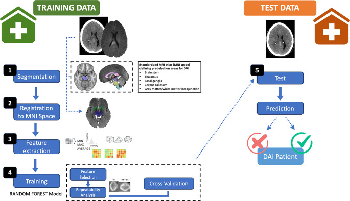
Methods: CT and MR imaging was performed in 42 patients suspicious of DAI due to the clinical state, and two control groups (n = 44;42). DAI was diagnosed by experienced neuroradiologists. Radiomics features were extracted using a standardized MRI-based atlas of the predilection areas for DAI. Different MRI and CT based models were trained and validated by five-fold cross validation. Diagnostic performance was compared to the reading of two experienced radiologists and further validated in an external test dataset.
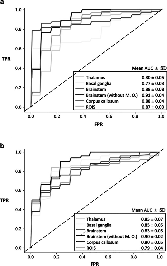
Results: The MRI and CT models showed significant differences in radiomics features between patients with DAI and controls. The developed MRI based random forest classifier yielded an accuracy of 80-90%. The best performing CT model yielded an accuracy of 88% in the training data and 70% in the external test data. The results were comparable to conventional image analysis which achieved an accuracy of 70-81% for CT-based diagnosis.
Conclusion: MRI- and CT-based radiomics analysis is feasible for the assessment of DAI. The radiomics classifier achieved equivalent performance rates as visual radiological image diagnosis. Especially a radiomics based CT classifier can be of clinical value as a screening and AI-based decision support tool for patients with TBI.
期刊介绍:
Clinical Neuroradiology provides current information, original contributions, and reviews in the field of neuroradiology. An interdisciplinary approach is accomplished by diagnostic and therapeutic contributions related to associated subjects.
The international coverage and relevance of the journal is underlined by its being the official journal of the German, Swiss, and Austrian Societies of Neuroradiology.
 求助内容:
求助内容: 应助结果提醒方式:


