Samantha J. Knott, Trisha Tucholski, Harini Josyer, David Inman, Andreas Friedl, Yanlong Zhu, Ying Ge* and Suzanne M. Ponik*,
{"title":"Deciphering Proteoform Landscape of Mammary Carcinoma by Top-Down Proteomics","authors":"Samantha J. Knott, Trisha Tucholski, Harini Josyer, David Inman, Andreas Friedl, Yanlong Zhu, Ying Ge* and Suzanne M. Ponik*, ","doi":"10.1021/acs.jproteome.4c0104410.1021/acs.jproteome.4c01044","DOIUrl":null,"url":null,"abstract":"<p >Defining the proteoform landscape of breast cancer can provide unique insights into the signaling pathways driving disease progression. While bottom-up proteomics has been utilized to profile breast cancer, it lacks the ability to capture intact proteoforms that may underpin the disease. Top-down proteomics is ideally suited to characterize intact proteoforms; however, most top-down proteomics studies have been limited to low molecular weight (MW) proteins (<50 kDa). Herein, we employed a two-dimensional (2D) liquid chromatography combining size exclusion chromatography (SEC) with reverse phase chromatography (RPC) followed by high-resolution mass spectrometry (MS) to expand the coverage for high MW proteoforms. Using this 2D-SEC-RPC-MS approach, we observed a 5-fold increase in the detection of high MW proteoforms (>50 kDa) compared to the conventional 1D-RPC-MS. SEC separation significantly enhanced the detection of high MW proteoforms (>104 kDa), including intermediate filament proteins, vimentin and keratins. Based on accurate mass measurements and MS/MS data, we identified 775 proteoforms from both TFA and HEPES extracts and detected PTMs, such as acetylation, glutathionylation, and myristoylation. Pathway analysis uncovered many proteoforms involved in processes dysregulated in cancer progression. Overall, our findings illustrate the power of top-down proteomics in defining the proteoform landscape of breast carcinoma.</p>","PeriodicalId":48,"journal":{"name":"Journal of Proteome Research","volume":"24 3","pages":"1425–1438 1425–1438"},"PeriodicalIF":3.6000,"publicationDate":"2025-02-12","publicationTypes":"Journal Article","fieldsOfStudy":null,"isOpenAccess":false,"openAccessPdf":"","citationCount":"0","resultStr":null,"platform":"Semanticscholar","paperid":null,"PeriodicalName":"Journal of Proteome Research","FirstCategoryId":"99","ListUrlMain":"https://pubs.acs.org/doi/10.1021/acs.jproteome.4c01044","RegionNum":2,"RegionCategory":"生物学","ArticlePicture":[],"TitleCN":null,"AbstractTextCN":null,"PMCID":null,"EPubDate":"","PubModel":"","JCR":"Q1","JCRName":"BIOCHEMICAL RESEARCH METHODS","Score":null,"Total":0}
引用次数: 0
Abstract
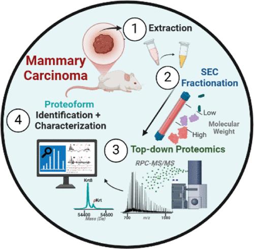
Defining the proteoform landscape of breast cancer can provide unique insights into the signaling pathways driving disease progression. While bottom-up proteomics has been utilized to profile breast cancer, it lacks the ability to capture intact proteoforms that may underpin the disease. Top-down proteomics is ideally suited to characterize intact proteoforms; however, most top-down proteomics studies have been limited to low molecular weight (MW) proteins (<50 kDa). Herein, we employed a two-dimensional (2D) liquid chromatography combining size exclusion chromatography (SEC) with reverse phase chromatography (RPC) followed by high-resolution mass spectrometry (MS) to expand the coverage for high MW proteoforms. Using this 2D-SEC-RPC-MS approach, we observed a 5-fold increase in the detection of high MW proteoforms (>50 kDa) compared to the conventional 1D-RPC-MS. SEC separation significantly enhanced the detection of high MW proteoforms (>104 kDa), including intermediate filament proteins, vimentin and keratins. Based on accurate mass measurements and MS/MS data, we identified 775 proteoforms from both TFA and HEPES extracts and detected PTMs, such as acetylation, glutathionylation, and myristoylation. Pathway analysis uncovered many proteoforms involved in processes dysregulated in cancer progression. Overall, our findings illustrate the power of top-down proteomics in defining the proteoform landscape of breast carcinoma.
期刊介绍:
Journal of Proteome Research publishes content encompassing all aspects of global protein analysis and function, including the dynamic aspects of genomics, spatio-temporal proteomics, metabonomics and metabolomics, clinical and agricultural proteomics, as well as advances in methodology including bioinformatics. The theme and emphasis is on a multidisciplinary approach to the life sciences through the synergy between the different types of "omics".
 求助内容:
求助内容: 应助结果提醒方式:


