Impact of loading, heart rate, and short episodes of ischaemia on myocardial stiffness assessed using shear wave elastography in an open-chest animal model.
Eric Saloux, Christophe Simard, Pauline Ruello, Adrien Lemaitre, Amir Hodzic, Alexandre Lebrun, Pierre-Antoine Dupont, Christophe Tribouilloy, Hélène Eltchaninoff, Morgane Le Garec, Christophe Fraschini, Vladimir Saplacan, Alain Manrique
{"title":"Impact of loading, heart rate, and short episodes of ischaemia on myocardial stiffness assessed using shear wave elastography in an open-chest animal model.","authors":"Eric Saloux, Christophe Simard, Pauline Ruello, Adrien Lemaitre, Amir Hodzic, Alexandre Lebrun, Pierre-Antoine Dupont, Christophe Tribouilloy, Hélène Eltchaninoff, Morgane Le Garec, Christophe Fraschini, Vladimir Saplacan, Alain Manrique","doi":"10.1093/ehjimp/qyaf015","DOIUrl":null,"url":null,"abstract":"<p><strong>Aims: </strong>Shear wave elastography (SWE) is a new promising ultrasound modality that enables non-invasive measurement of the dynamic myocardial stiffness. The impact of varying physiological conditions on SWE measurement of left ventricular (LV) myocardial stiffness remains poorly investigated.</p><p><strong>Methods and results: </strong>Nineteen sheep were evaluated during open-chest surgery. Epicardial multiframe SWE acquisitions were performed in short-axis view simultaneously with haemodynamic acquisitions during inferior vena cava occlusion, aortic clamping, atrial pacing, and ischaemia-reperfusion. The cyclic variation in the median value of LV myocardial stiffness ranged from 1.1 m/s in diastole (C<sub>min</sub>) to 2.4 m/s in systole (C<sub>max</sub>). At steady state, intra-animal reproducibility was good for C<sub>min</sub> [intraclass correlation coefficient ICC = 0.77 (0.54, 0.90), <i>P</i> < 0.001] and C<sub>max</sub> [ICC = 0.92 (0.84, 0.96), <i>P</i> < 0.001]. C<sub>min</sub> was independent of loading conditions, heart rate, and short 15-minute episodes of ischaemia and reperfusion. C<sub>max</sub> was independent of loading conditions and moderate increase in heart rate but decreased significantly during ischaemia and reperfusion. Compared with baseline, percentage changes in C<sub>max</sub> was correlated to percentage changes in dP/dt<sub>max</sub> (<i>R</i> = 0.47, <i>P</i> = 0.001) and in LV systolic pressure (<i>R</i> = 0.35, <i>P</i> = 0.013) and SW (<i>R</i> = 0.31, <i>P</i> = 0.026).</p><p><strong>Conclusion: </strong>In this study, LV diastolic myocardial stiffness C<sub>min</sub> assessed using SWE demonstrated the characteristics of a potentially useful clinical marker of LV diastolic function linked to the intrinsic elastic properties of the myocardium, whereas C<sub>max</sub> was an indicator of LV contractility.</p>","PeriodicalId":94317,"journal":{"name":"European heart journal. Imaging methods and practice","volume":"3 1","pages":"qyaf015"},"PeriodicalIF":0.0000,"publicationDate":"2025-02-10","publicationTypes":"Journal Article","fieldsOfStudy":null,"isOpenAccess":false,"openAccessPdf":"https://www.ncbi.nlm.nih.gov/pmc/articles/PMC11879029/pdf/","citationCount":"0","resultStr":null,"platform":"Semanticscholar","paperid":null,"PeriodicalName":"European heart journal. Imaging methods and practice","FirstCategoryId":"1085","ListUrlMain":"https://doi.org/10.1093/ehjimp/qyaf015","RegionNum":0,"RegionCategory":null,"ArticlePicture":[],"TitleCN":null,"AbstractTextCN":null,"PMCID":null,"EPubDate":"2025/1/1 0:00:00","PubModel":"eCollection","JCR":"","JCRName":"","Score":null,"Total":0}
引用次数: 0
Abstract
Aims: Shear wave elastography (SWE) is a new promising ultrasound modality that enables non-invasive measurement of the dynamic myocardial stiffness. The impact of varying physiological conditions on SWE measurement of left ventricular (LV) myocardial stiffness remains poorly investigated.
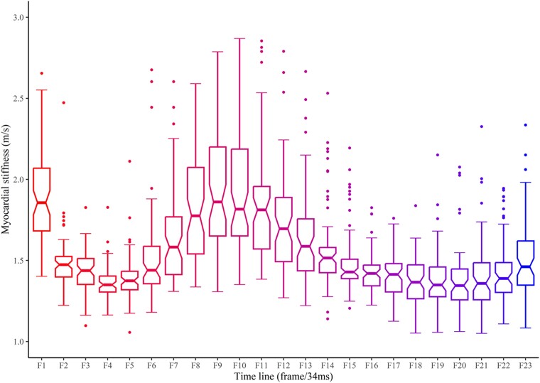
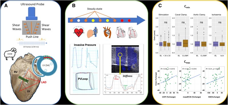
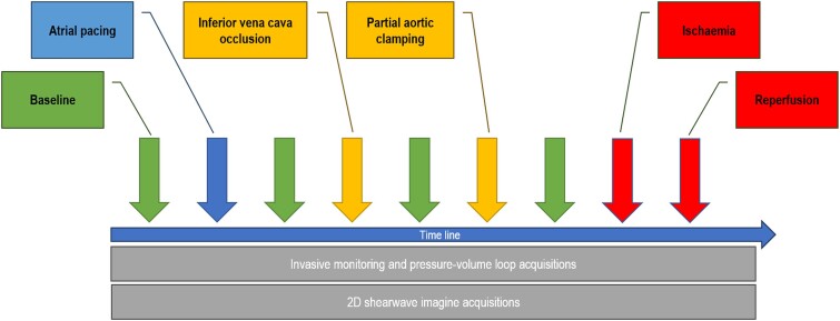
Methods and results: Nineteen sheep were evaluated during open-chest surgery. Epicardial multiframe SWE acquisitions were performed in short-axis view simultaneously with haemodynamic acquisitions during inferior vena cava occlusion, aortic clamping, atrial pacing, and ischaemia-reperfusion. The cyclic variation in the median value of LV myocardial stiffness ranged from 1.1 m/s in diastole (Cmin) to 2.4 m/s in systole (Cmax). At steady state, intra-animal reproducibility was good for Cmin [intraclass correlation coefficient ICC = 0.77 (0.54, 0.90), P < 0.001] and Cmax [ICC = 0.92 (0.84, 0.96), P < 0.001]. Cmin was independent of loading conditions, heart rate, and short 15-minute episodes of ischaemia and reperfusion. Cmax was independent of loading conditions and moderate increase in heart rate but decreased significantly during ischaemia and reperfusion. Compared with baseline, percentage changes in Cmax was correlated to percentage changes in dP/dtmax (R = 0.47, P = 0.001) and in LV systolic pressure (R = 0.35, P = 0.013) and SW (R = 0.31, P = 0.026).
Conclusion: In this study, LV diastolic myocardial stiffness Cmin assessed using SWE demonstrated the characteristics of a potentially useful clinical marker of LV diastolic function linked to the intrinsic elastic properties of the myocardium, whereas Cmax was an indicator of LV contractility.

 求助内容:
求助内容: 应助结果提醒方式:


