Ahmet Baytok, Gökhan Ecer, Mehmet Balasar, Mustafa Koplay
{"title":"Computed tomography and magnetic resonance imaging characteristics of renal cell carcinoma: Differences between subtypes and clinical evaluation.","authors":"Ahmet Baytok, Gökhan Ecer, Mehmet Balasar, Mustafa Koplay","doi":"10.25259/JCIS_160_2024","DOIUrl":null,"url":null,"abstract":"<p><p>This review discusses the evaluation of renal cell carcinoma (RCC) subtypes using computed tomography (CT) and magnetic resonance imaging (MRI). RCC is a malignancy with different histopathological subtypes, constituting approximately 90% of adult kidney tumors. It has been reported that these subtypes show significant differences in terms of clinical behavior, treatment response, and prognosis. In the study, CT and MRI findings of subtypes such as clear cell RCC (ccRCC), papillary RCC (pRCC), chromophobe RCC (chRCC), medullary RCC (mRCC), collecting duct RCC (cdRCC), and multiloculated cystic RCC (mcRCC) were compared. It was stated that CT is the first-choice imaging method in the staging and surgical planning of RCC and provides detailed information about the tumor size, vascularity, and metastatic spread. On the other hand, it has been emphasized that MRI allows better characterization of RCC subtypes with its soft-tissue resolution and contrast agent usage advantage. The study draws attention to the different imaging features of each subtype and details the role of these findings in the clinical decision-making process. It has been stated that ccRCC exhibits intense contrast enhancement and rapid washout pattern in the corticomedullary phase on CT and appears hyperintense on T2A and hypointense on T1 weighted imaging (T1A) on MRI. It has been stated that pRCC has hypovascular features, has lower contrast enhancement, and has homogeneous borders. It has been stated that chRCC has a less vascular structure and exhibits moderate contrast enhancement in the corticomedullary phase. It has been reported that mRCC has invasive features and is usually diagnosed at an advanced stage while cdRCC has a very aggressive clinical course. It has been stated that mcRCC contains distinct cystic areas between the septa, has a well-circumscribed structure, and generally has a low malignancy potential. As a result, it has been stated that detailed evaluation of CT and MRI findings of RCC subtypes plays a critical role in the diagnosis, treatment, and prognosis of these subtypes. It has been emphasized that the findings presented in this study will contribute to the development of more targeted treatment approaches in RCC management.</p>","PeriodicalId":15512,"journal":{"name":"Journal of Clinical Imaging Science","volume":"15 ","pages":"10"},"PeriodicalIF":1.3000,"publicationDate":"2025-02-25","publicationTypes":"Journal Article","fieldsOfStudy":null,"isOpenAccess":false,"openAccessPdf":"https://www.ncbi.nlm.nih.gov/pmc/articles/PMC11878704/pdf/","citationCount":"0","resultStr":null,"platform":"Semanticscholar","paperid":null,"PeriodicalName":"Journal of Clinical Imaging Science","FirstCategoryId":"1085","ListUrlMain":"https://doi.org/10.25259/JCIS_160_2024","RegionNum":0,"RegionCategory":null,"ArticlePicture":[],"TitleCN":null,"AbstractTextCN":null,"PMCID":null,"EPubDate":"2025/1/1 0:00:00","PubModel":"eCollection","JCR":"Q3","JCRName":"RADIOLOGY, NUCLEAR MEDICINE & MEDICAL IMAGING","Score":null,"Total":0}
引用次数: 0
Abstract
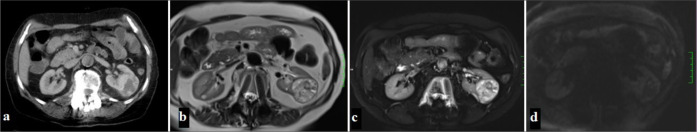
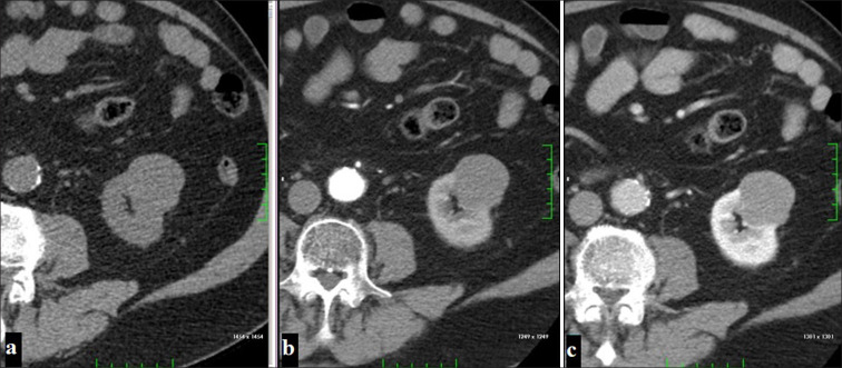
This review discusses the evaluation of renal cell carcinoma (RCC) subtypes using computed tomography (CT) and magnetic resonance imaging (MRI). RCC is a malignancy with different histopathological subtypes, constituting approximately 90% of adult kidney tumors. It has been reported that these subtypes show significant differences in terms of clinical behavior, treatment response, and prognosis. In the study, CT and MRI findings of subtypes such as clear cell RCC (ccRCC), papillary RCC (pRCC), chromophobe RCC (chRCC), medullary RCC (mRCC), collecting duct RCC (cdRCC), and multiloculated cystic RCC (mcRCC) were compared. It was stated that CT is the first-choice imaging method in the staging and surgical planning of RCC and provides detailed information about the tumor size, vascularity, and metastatic spread. On the other hand, it has been emphasized that MRI allows better characterization of RCC subtypes with its soft-tissue resolution and contrast agent usage advantage. The study draws attention to the different imaging features of each subtype and details the role of these findings in the clinical decision-making process. It has been stated that ccRCC exhibits intense contrast enhancement and rapid washout pattern in the corticomedullary phase on CT and appears hyperintense on T2A and hypointense on T1 weighted imaging (T1A) on MRI. It has been stated that pRCC has hypovascular features, has lower contrast enhancement, and has homogeneous borders. It has been stated that chRCC has a less vascular structure and exhibits moderate contrast enhancement in the corticomedullary phase. It has been reported that mRCC has invasive features and is usually diagnosed at an advanced stage while cdRCC has a very aggressive clinical course. It has been stated that mcRCC contains distinct cystic areas between the septa, has a well-circumscribed structure, and generally has a low malignancy potential. As a result, it has been stated that detailed evaluation of CT and MRI findings of RCC subtypes plays a critical role in the diagnosis, treatment, and prognosis of these subtypes. It has been emphasized that the findings presented in this study will contribute to the development of more targeted treatment approaches in RCC management.
期刊介绍:
The Journal of Clinical Imaging Science (JCIS) is an open access peer-reviewed journal committed to publishing high-quality articles in the field of Imaging Science. The journal aims to present Imaging Science and relevant clinical information in an understandable and useful format. The journal is owned and published by the Scientific Scholar. Audience Our audience includes Radiologists, Researchers, Clinicians, medical professionals and students. Review process JCIS has a highly rigorous peer-review process that makes sure that manuscripts are scientifically accurate, relevant, novel and important. Authors disclose all conflicts, affiliations and financial associations such that the published content is not biased.

 求助内容:
求助内容: 应助结果提醒方式:


