{"title":"Basic verification of myocardial extracellular volume quantification by prototype photon-counting detector computed tomography: A phantom study.","authors":"Seitaro Oda, Yoshinori Funama, Shinichi Kojima, Kazuma Yokoi, Isao Takahashi, Yuko Aoki, Taiga Goto, Kana Tanaka, Fuyuhiko Teramoto, Masafumi Kidoh, Yasunori Nagayama, Takeshi Nakaura, Toshinori Hirai","doi":"10.25259/JCIS_157_2024","DOIUrl":null,"url":null,"abstract":"<p><strong>Objectives: </strong>This study aimed to investigate the accuracy of myocardial extracellular volume (ECV) quantification using a prototype photon-counting detector (PCD) computed tomography (CT) and examine the association between radiation dose and spectral image settings.</p><p><strong>Material and methods: </strong>A multi-energy CT phantom that simulated the blood pool and myocardium was used. The tube voltage was set at 120 kVp and three types of tube current-time products (105, 150, and 300 mAs) were applied for pre- and post-contrast scans. Virtual monoenergetic images (VMIs) at 50-100 keV were reconstructed. The ECV value was calculated from the CT numbers between pre-contrast and post-contrast. We compared the accuracy of ECV values at each VMI level.</p><p><strong>Results: </strong>Each radiation dose setting demonstrated a small but significant difference in ECV values at each keV level. ECV was overestimated at higher keV in all radiation dose settings. A significant difference in ECV value variabilities was found among keV levels in all three radiation dose settings, with higher keV exhibiting greater variability. The variation was particularly large in the low-dose setting. The residual values were significantly larger at higher keV levels in all radiation dose settings. The residual values were smaller at 50 and 60 keV with no significant difference in 150- and 300-mAs settings.</p><p><strong>Conclusion: </strong>Setting appropriate VMI keV and radiation dose settings was necessary when quantifying myocardial ECV with PCD-CT because the keV levels caused differences in the quantification value and measurement variation.</p>","PeriodicalId":15512,"journal":{"name":"Journal of Clinical Imaging Science","volume":"15 ","pages":"8"},"PeriodicalIF":1.3000,"publicationDate":"2025-02-11","publicationTypes":"Journal Article","fieldsOfStudy":null,"isOpenAccess":false,"openAccessPdf":"https://www.ncbi.nlm.nih.gov/pmc/articles/PMC11878665/pdf/","citationCount":"0","resultStr":null,"platform":"Semanticscholar","paperid":null,"PeriodicalName":"Journal of Clinical Imaging Science","FirstCategoryId":"1085","ListUrlMain":"https://doi.org/10.25259/JCIS_157_2024","RegionNum":0,"RegionCategory":null,"ArticlePicture":[],"TitleCN":null,"AbstractTextCN":null,"PMCID":null,"EPubDate":"2025/1/1 0:00:00","PubModel":"eCollection","JCR":"Q3","JCRName":"RADIOLOGY, NUCLEAR MEDICINE & MEDICAL IMAGING","Score":null,"Total":0}
引用次数: 0
Abstract
Objectives: This study aimed to investigate the accuracy of myocardial extracellular volume (ECV) quantification using a prototype photon-counting detector (PCD) computed tomography (CT) and examine the association between radiation dose and spectral image settings.
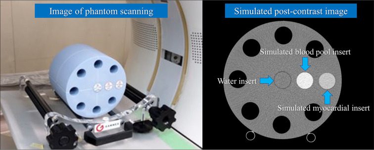
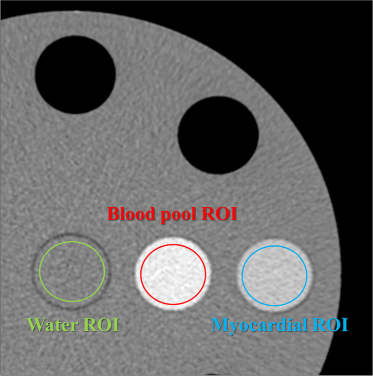
Material and methods: A multi-energy CT phantom that simulated the blood pool and myocardium was used. The tube voltage was set at 120 kVp and three types of tube current-time products (105, 150, and 300 mAs) were applied for pre- and post-contrast scans. Virtual monoenergetic images (VMIs) at 50-100 keV were reconstructed. The ECV value was calculated from the CT numbers between pre-contrast and post-contrast. We compared the accuracy of ECV values at each VMI level.
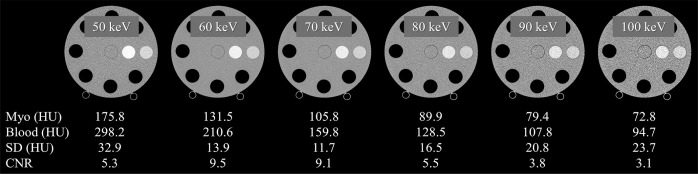
Results: Each radiation dose setting demonstrated a small but significant difference in ECV values at each keV level. ECV was overestimated at higher keV in all radiation dose settings. A significant difference in ECV value variabilities was found among keV levels in all three radiation dose settings, with higher keV exhibiting greater variability. The variation was particularly large in the low-dose setting. The residual values were significantly larger at higher keV levels in all radiation dose settings. The residual values were smaller at 50 and 60 keV with no significant difference in 150- and 300-mAs settings.
Conclusion: Setting appropriate VMI keV and radiation dose settings was necessary when quantifying myocardial ECV with PCD-CT because the keV levels caused differences in the quantification value and measurement variation.
期刊介绍:
The Journal of Clinical Imaging Science (JCIS) is an open access peer-reviewed journal committed to publishing high-quality articles in the field of Imaging Science. The journal aims to present Imaging Science and relevant clinical information in an understandable and useful format. The journal is owned and published by the Scientific Scholar. Audience Our audience includes Radiologists, Researchers, Clinicians, medical professionals and students. Review process JCIS has a highly rigorous peer-review process that makes sure that manuscripts are scientifically accurate, relevant, novel and important. Authors disclose all conflicts, affiliations and financial associations such that the published content is not biased.

 求助内容:
求助内容: 应助结果提醒方式:


