Hedvig Elfving, Hui Yu, Kaleab Kassete Fessehatsion, Hans Brunnström, Johan Botling, Miklos Gulyas, Max Backman, Amanda Lindberg, Carina Strell, Patrick Micke
{"title":"Spatial distribution of tertiary lymphoid structures in the molecular and clinical context of non-small cell lung cancer.","authors":"Hedvig Elfving, Hui Yu, Kaleab Kassete Fessehatsion, Hans Brunnström, Johan Botling, Miklos Gulyas, Max Backman, Amanda Lindberg, Carina Strell, Patrick Micke","doi":"10.1007/s13402-025-01052-x","DOIUrl":null,"url":null,"abstract":"<p><strong>Introduction: </strong>Tertiary lymphoid structures (TLS) are lymphocyte aggregates resembling secondary lymphoid organs and are pivotal in cancer immunity. The ambiguous morphological definition of TLS makes it challenging to ascertain their clinical impact on patient survival and response to immunotherapy.</p><p><strong>Objectives: </strong>This study aimed to characterize TLS in hematoxylin-eosin tissue sections from lung cancer patients, assessing their occurrence in relation to the local immune environment, mutational background, and patient outcome.</p><p><strong>Methods: </strong>Two pathologists evaluated one whole tissue section from resection specimens of 680 NSCLC patients. TLS were spatially quantified within the tumor area or periphery and further categorized based on the presence of germinal centers (mature TLS). Metrics were integrated with immune cell counts, genomic and transcriptomic data, and correlated with clinical parameters.</p><p><strong>Results: </strong>TLS were present in 86% of 536 evaluable cases, predominantly in the tumor periphery, with a median of eight TLS per case. Mature TLS were found in 24% of cases. TLS presence correlated positively with increased plasma cell (CD138+) and lymphocytic cell (CD3+, CD8+, FOXP3+) infiltration. Tumors with higher tumor mutational burden exhibited higher numbers of peripheral TLS. The overall TLS quantity was independently associated with improved patient survival, irrespective of TLS maturation status. This prognostic association held true for peripheral TLS but not for tumor TLS.</p><p><strong>Conclusion: </strong>TLS in NSCLC is common and their correlation with a specific immune phenotype suggests biological relevance in the local immune reaction. The prognostic significance of this scoring system on routine hematoxylin-eosin sections has the potential to augment diagnostic algorithms for NSCLC patients.</p>","PeriodicalId":49223,"journal":{"name":"Cellular Oncology","volume":" ","pages":"801-813"},"PeriodicalIF":4.8000,"publicationDate":"2025-06-01","publicationTypes":"Journal Article","fieldsOfStudy":null,"isOpenAccess":false,"openAccessPdf":"https://www.ncbi.nlm.nih.gov/pmc/articles/PMC12119696/pdf/","citationCount":"0","resultStr":null,"platform":"Semanticscholar","paperid":null,"PeriodicalName":"Cellular Oncology","FirstCategoryId":"3","ListUrlMain":"https://doi.org/10.1007/s13402-025-01052-x","RegionNum":2,"RegionCategory":"医学","ArticlePicture":[],"TitleCN":null,"AbstractTextCN":null,"PMCID":null,"EPubDate":"2025/3/3 0:00:00","PubModel":"Epub","JCR":"Q2","JCRName":"CELL BIOLOGY","Score":null,"Total":0}
引用次数: 0
Abstract
Introduction: Tertiary lymphoid structures (TLS) are lymphocyte aggregates resembling secondary lymphoid organs and are pivotal in cancer immunity. The ambiguous morphological definition of TLS makes it challenging to ascertain their clinical impact on patient survival and response to immunotherapy.
Objectives: This study aimed to characterize TLS in hematoxylin-eosin tissue sections from lung cancer patients, assessing their occurrence in relation to the local immune environment, mutational background, and patient outcome.
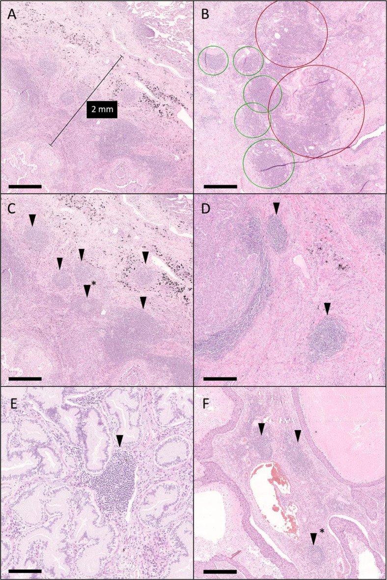
Methods: Two pathologists evaluated one whole tissue section from resection specimens of 680 NSCLC patients. TLS were spatially quantified within the tumor area or periphery and further categorized based on the presence of germinal centers (mature TLS). Metrics were integrated with immune cell counts, genomic and transcriptomic data, and correlated with clinical parameters.
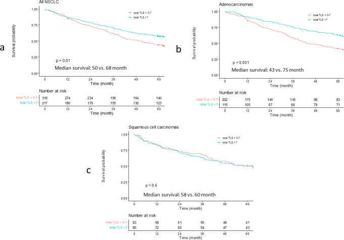
Results: TLS were present in 86% of 536 evaluable cases, predominantly in the tumor periphery, with a median of eight TLS per case. Mature TLS were found in 24% of cases. TLS presence correlated positively with increased plasma cell (CD138+) and lymphocytic cell (CD3+, CD8+, FOXP3+) infiltration. Tumors with higher tumor mutational burden exhibited higher numbers of peripheral TLS. The overall TLS quantity was independently associated with improved patient survival, irrespective of TLS maturation status. This prognostic association held true for peripheral TLS but not for tumor TLS.
Conclusion: TLS in NSCLC is common and their correlation with a specific immune phenotype suggests biological relevance in the local immune reaction. The prognostic significance of this scoring system on routine hematoxylin-eosin sections has the potential to augment diagnostic algorithms for NSCLC patients.
期刊介绍:
The Official Journal of the International Society for Cellular Oncology
Focuses on translational research
Addresses the conversion of cell biology to clinical applications
Cellular Oncology publishes scientific contributions from various biomedical and clinical disciplines involved in basic and translational cancer research on the cell and tissue level, technical and bioinformatics developments in this area, and clinical applications. This includes a variety of fields like genome technology, micro-arrays and other high-throughput techniques, genomic instability, SNP, DNA methylation, signaling pathways, DNA organization, (sub)microscopic imaging, proteomics, bioinformatics, functional effects of genomics, drug design and development, molecular diagnostics and targeted cancer therapies, genotype-phenotype interactions.
A major goal is to translate the latest developments in these fields from the research laboratory into routine patient management. To this end Cellular Oncology forms a platform of scientific information exchange between molecular biologists and geneticists, technical developers, pathologists, (medical) oncologists and other clinicians involved in the management of cancer patients.
In vitro studies are preferentially supported by validations in tumor tissue with clinicopathological associations.

 求助内容:
求助内容: 应助结果提醒方式:


