Parsa Khalafi, Soroush Morsali, Sana Hamidi, Hamidreza Ashayeri, Navid Sobhi, Siamak Pedrammehr, Ali Jafarizadeh
{"title":"Artificial intelligence in stroke risk assessment and management via retinal imaging.","authors":"Parsa Khalafi, Soroush Morsali, Sana Hamidi, Hamidreza Ashayeri, Navid Sobhi, Siamak Pedrammehr, Ali Jafarizadeh","doi":"10.3389/fncom.2025.1490603","DOIUrl":null,"url":null,"abstract":"<p><p>Retinal imaging, used for assessing stroke-related retinal changes, is a non-invasive and cost-effective method that can be enhanced by machine learning and deep learning algorithms, showing promise in early disease detection, severity grading, and prognostic evaluation in stroke patients. This review explores the role of artificial intelligence (AI) in stroke patient care, focusing on retinal imaging integration into clinical workflows. Retinal imaging has revealed several microvascular changes, including a decrease in the central retinal artery diameter and an increase in the central retinal vein diameter, both of which are associated with lacunar stroke and intracranial hemorrhage. Additionally, microvascular changes, such as arteriovenous nicking, increased vessel tortuosity, enhanced arteriolar light reflex, decreased retinal fractals, and thinning of retinal nerve fiber layer are also reported to be associated with higher stroke risk. AI models, such as Xception and EfficientNet, have demonstrated accuracy comparable to traditional stroke risk scoring systems in predicting stroke risk. For stroke diagnosis, models like Inception, ResNet, and VGG, alongside machine learning classifiers, have shown high efficacy in distinguishing stroke patients from healthy individuals using retinal imaging. Moreover, a random forest model effectively distinguished between ischemic and hemorrhagic stroke subtypes based on retinal features, showing superior predictive performance compared to traditional clinical characteristics. Additionally, a support vector machine model has achieved high classification accuracy in assessing pial collateral status. Despite this advancements, challenges such as the lack of standardized protocols for imaging modalities, hesitance in trusting AI-generated predictions, insufficient integration of retinal imaging data with electronic health records, the need for validation across diverse populations, and ethical and regulatory concerns persist. Future efforts must focus on validating AI models across diverse populations, ensuring algorithm transparency, and addressing ethical and regulatory issues to enable broader implementation. Overcoming these barriers will be essential for translating this technology into personalized stroke care and improving patient outcomes.</p>","PeriodicalId":12363,"journal":{"name":"Frontiers in Computational Neuroscience","volume":"19 ","pages":"1490603"},"PeriodicalIF":2.3000,"publicationDate":"2025-02-17","publicationTypes":"Journal Article","fieldsOfStudy":null,"isOpenAccess":false,"openAccessPdf":"https://www.ncbi.nlm.nih.gov/pmc/articles/PMC11872910/pdf/","citationCount":"0","resultStr":null,"platform":"Semanticscholar","paperid":null,"PeriodicalName":"Frontiers in Computational Neuroscience","FirstCategoryId":"3","ListUrlMain":"https://doi.org/10.3389/fncom.2025.1490603","RegionNum":4,"RegionCategory":"医学","ArticlePicture":[],"TitleCN":null,"AbstractTextCN":null,"PMCID":null,"EPubDate":"2025/1/1 0:00:00","PubModel":"eCollection","JCR":"Q2","JCRName":"MATHEMATICAL & COMPUTATIONAL BIOLOGY","Score":null,"Total":0}
引用次数: 0
Abstract
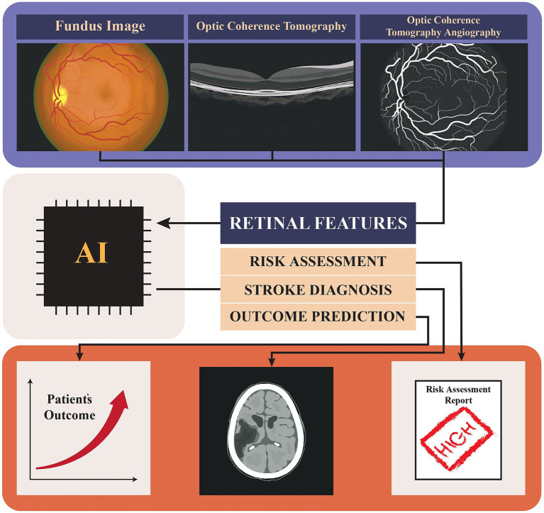
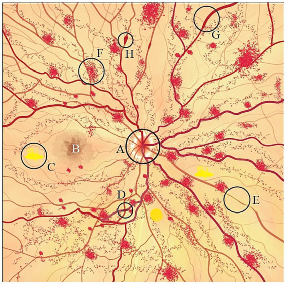
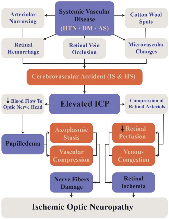
Retinal imaging, used for assessing stroke-related retinal changes, is a non-invasive and cost-effective method that can be enhanced by machine learning and deep learning algorithms, showing promise in early disease detection, severity grading, and prognostic evaluation in stroke patients. This review explores the role of artificial intelligence (AI) in stroke patient care, focusing on retinal imaging integration into clinical workflows. Retinal imaging has revealed several microvascular changes, including a decrease in the central retinal artery diameter and an increase in the central retinal vein diameter, both of which are associated with lacunar stroke and intracranial hemorrhage. Additionally, microvascular changes, such as arteriovenous nicking, increased vessel tortuosity, enhanced arteriolar light reflex, decreased retinal fractals, and thinning of retinal nerve fiber layer are also reported to be associated with higher stroke risk. AI models, such as Xception and EfficientNet, have demonstrated accuracy comparable to traditional stroke risk scoring systems in predicting stroke risk. For stroke diagnosis, models like Inception, ResNet, and VGG, alongside machine learning classifiers, have shown high efficacy in distinguishing stroke patients from healthy individuals using retinal imaging. Moreover, a random forest model effectively distinguished between ischemic and hemorrhagic stroke subtypes based on retinal features, showing superior predictive performance compared to traditional clinical characteristics. Additionally, a support vector machine model has achieved high classification accuracy in assessing pial collateral status. Despite this advancements, challenges such as the lack of standardized protocols for imaging modalities, hesitance in trusting AI-generated predictions, insufficient integration of retinal imaging data with electronic health records, the need for validation across diverse populations, and ethical and regulatory concerns persist. Future efforts must focus on validating AI models across diverse populations, ensuring algorithm transparency, and addressing ethical and regulatory issues to enable broader implementation. Overcoming these barriers will be essential for translating this technology into personalized stroke care and improving patient outcomes.
期刊介绍:
Frontiers in Computational Neuroscience is a first-tier electronic journal devoted to promoting theoretical modeling of brain function and fostering interdisciplinary interactions between theoretical and experimental neuroscience. Progress in understanding the amazing capabilities of the brain is still limited, and we believe that it will only come with deep theoretical thinking and mutually stimulating cooperation between different disciplines and approaches. We therefore invite original contributions on a wide range of topics that present the fruits of such cooperation, or provide stimuli for future alliances. We aim to provide an interactive forum for cutting-edge theoretical studies of the nervous system, and for promulgating the best theoretical research to the broader neuroscience community. Models of all styles and at all levels are welcome, from biophysically motivated realistic simulations of neurons and synapses to high-level abstract models of inference and decision making. While the journal is primarily focused on theoretically based and driven research, we welcome experimental studies that validate and test theoretical conclusions.
Also: comp neuro
 求助内容:
求助内容: 应助结果提醒方式:


