Mengfei Liu, Zifan Qi, Ren Zhou, Chuanhai Guo, Anxiang Liu, Haijun Yang, Fenglei Li, Liping Duan, Lin Shen, Qi Wu, Zhen Liu, Yaqi Pan, Fangfang Liu, Ying Liu, Huanyu Chen, Zhe Hu, Hong Cai, Zhonghu He, Yang Ke
{"title":"An Image-Based Model for Assisting in Diagnosing Malignant Esophageal Lesions During Lugol Chromoendoscopic Examination.","authors":"Mengfei Liu, Zifan Qi, Ren Zhou, Chuanhai Guo, Anxiang Liu, Haijun Yang, Fenglei Li, Liping Duan, Lin Shen, Qi Wu, Zhen Liu, Yaqi Pan, Fangfang Liu, Ying Liu, Huanyu Chen, Zhe Hu, Hong Cai, Zhonghu He, Yang Ke","doi":"10.14309/ctg.0000000000000835","DOIUrl":null,"url":null,"abstract":"<p><strong>Introduction: </strong>Image-based diagnostic tools that aid endoscopists to biopsy putative esophageal malignant lesions are essential for ensuring the standardization and quality of Lugol chromoendoscopy. But there is no such model available yet.</p><p><strong>Methods: </strong>We developed a diagnostic model using endoscopic Lugol-unstained lesions (LULs) features and baseline data from 1,099 individuals enrolled from a large-scale population-based ESCC screening cohort. Six hundred three participants from a clinical outpatient cohort were included as the external validation set. High-grade intraepithelial neoplasia and above lesions identified at baseline or within 1 year after screening were defined as outcome. The final model was determined using logistic regression analysis by the Akaike information criterion.</p><p><strong>Results: </strong>The optimal diagnostic model contained the size, irregularity, sharp border of LUL, age, and body mass index of the participant, with the area under the curve of 0.83 (95% confidence interval [CI]: 0.78-0.87) in the development set, 0.81 (95% CI: 0.77-0.86) in the internal validation set, and 0.87 (95% CI: 0.84-0.90) in the external set. This model stratified individuals with LULs into low-risk, moderate-risk, and high-risk groups based on tertiles of predicted probabilities. The high-risk group accounted for <40% participants but enriched 80.8% and 82.7% of high-grade intraepithelial neoplasia and above cases in the development and external validation sets, respectively, achieving detection ratios 16.2 and 11.0 times higher than the low-risk group.</p><p><strong>Discussion: </strong>Our model can help maintain consistency and accuracy in detecting esophageal malignancy through Lugol chromoendoscopy, particularly in primary healthcare units in high-risk rural areas.</p>","PeriodicalId":10278,"journal":{"name":"Clinical and Translational Gastroenterology","volume":" ","pages":"e00835"},"PeriodicalIF":3.0000,"publicationDate":"2025-05-01","publicationTypes":"Journal Article","fieldsOfStudy":null,"isOpenAccess":false,"openAccessPdf":"https://www.ncbi.nlm.nih.gov/pmc/articles/PMC12101928/pdf/","citationCount":"0","resultStr":null,"platform":"Semanticscholar","paperid":null,"PeriodicalName":"Clinical and Translational Gastroenterology","FirstCategoryId":"3","ListUrlMain":"https://doi.org/10.14309/ctg.0000000000000835","RegionNum":3,"RegionCategory":"医学","ArticlePicture":[],"TitleCN":null,"AbstractTextCN":null,"PMCID":null,"EPubDate":"","PubModel":"","JCR":"Q2","JCRName":"GASTROENTEROLOGY & HEPATOLOGY","Score":null,"Total":0}
引用次数: 0
Abstract
Introduction: Image-based diagnostic tools that aid endoscopists to biopsy putative esophageal malignant lesions are essential for ensuring the standardization and quality of Lugol chromoendoscopy. But there is no such model available yet.
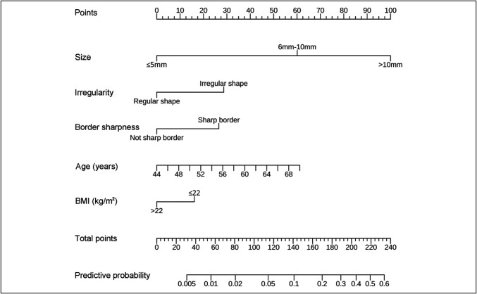
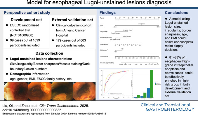
Methods: We developed a diagnostic model using endoscopic Lugol-unstained lesions (LULs) features and baseline data from 1,099 individuals enrolled from a large-scale population-based ESCC screening cohort. Six hundred three participants from a clinical outpatient cohort were included as the external validation set. High-grade intraepithelial neoplasia and above lesions identified at baseline or within 1 year after screening were defined as outcome. The final model was determined using logistic regression analysis by the Akaike information criterion.
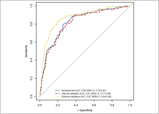
Results: The optimal diagnostic model contained the size, irregularity, sharp border of LUL, age, and body mass index of the participant, with the area under the curve of 0.83 (95% confidence interval [CI]: 0.78-0.87) in the development set, 0.81 (95% CI: 0.77-0.86) in the internal validation set, and 0.87 (95% CI: 0.84-0.90) in the external set. This model stratified individuals with LULs into low-risk, moderate-risk, and high-risk groups based on tertiles of predicted probabilities. The high-risk group accounted for <40% participants but enriched 80.8% and 82.7% of high-grade intraepithelial neoplasia and above cases in the development and external validation sets, respectively, achieving detection ratios 16.2 and 11.0 times higher than the low-risk group.
Discussion: Our model can help maintain consistency and accuracy in detecting esophageal malignancy through Lugol chromoendoscopy, particularly in primary healthcare units in high-risk rural areas.
期刊介绍:
Clinical and Translational Gastroenterology (CTG), published on behalf of the American College of Gastroenterology (ACG), is a peer-reviewed open access online journal dedicated to innovative clinical work in the field of gastroenterology and hepatology. CTG hopes to fulfill an unmet need for clinicians and scientists by welcoming novel cohort studies, early-phase clinical trials, qualitative and quantitative epidemiologic research, hypothesis-generating research, studies of novel mechanisms and methodologies including public health interventions, and integration of approaches across organs and disciplines. CTG also welcomes hypothesis-generating small studies, methods papers, and translational research with clear applications to human physiology or disease.
Colon and small bowel
Endoscopy and novel diagnostics
Esophagus
Functional GI disorders
Immunology of the GI tract
Microbiology of the GI tract
Inflammatory bowel disease
Pancreas and biliary tract
Liver
Pathology
Pediatrics
Preventative medicine
Nutrition/obesity
Stomach.
 求助内容:
求助内容: 应助结果提醒方式:


