Shi Shen, Benchao Shu, Yulin Xu, Heng Zhao, Yang Li, Yujie Li, Chuanchuan Zhuo, Naiqiang Zhuo
{"title":"Characterization and Biocompatibility Assessment of 3D-Printed HA/PCL Porous Bionic Bone Scaffold: <i>in Vitro</i> and <i>in Vivo</i> Evaluation.","authors":"Shi Shen, Benchao Shu, Yulin Xu, Heng Zhao, Yang Li, Yujie Li, Chuanchuan Zhuo, Naiqiang Zhuo","doi":"10.22540/JMNI-25-119","DOIUrl":null,"url":null,"abstract":"<p><strong>Objectives: </strong>This study aims to characterize a three-dimensional-printed hydroxyapatite (HA)/polycaprolactone (PCL) scaffold and assess its biocompatibility both <i>in vitro</i> and <i>in vivo</i>.</p><p><strong>Methods: </strong>A bionic, porous HA/PCL scaffold was fabricated using 3D printing, and its microstructure, porosity, hydrophilicity, and mechanical properties were evaluated through scanning electron microscopy and various assays. Bone marrow mesenchymal stem cells (BMSCs) and vascular endothelial progenitor cells (VEPCs) were co-cultured with the scaffold, and their proliferation and osteogenic differentiation were assessed using the Cell Counting Kit-8, ALP assays, and alizarin red staining. Osteogenic marker expression was analyzed via qRT-PCR. <i>In vivo</i> bone regeneration was evaluated through histological analysis of H&E and Masson's trichrome staining in a rat cranial defect model.</p><p><strong>Results: </strong>The average pore size of the scaffold was 462.00 ± 100.389 μm, with a porosity of 53%, a water absorption expansion rate of 5.10%, a contact angle of 94.55°, an elastic modulus of 53.82 MPa, and a compressive strength of 6.10 MPa. ALP activity and qRT-PCR analysis of osteogenic markers (BMP2, OCN, Runx2) showed significant upregulation in cells co-cultured with the scaffolds. In vivo experiments demonstrated enhanced bone regeneration and collagen deposition in the HA/PCL scaffold group.</p><p><strong>Conclusion: </strong>The results suggest that the HA/PCL scaffold promotes osteogenic differentiation and bone regeneration, making it suitable for bone tissue engineering applications.</p>","PeriodicalId":16430,"journal":{"name":"Journal of musculoskeletal & neuronal interactions","volume":"25 1","pages":"119-132"},"PeriodicalIF":1.6000,"publicationDate":"2025-03-01","publicationTypes":"Journal Article","fieldsOfStudy":null,"isOpenAccess":false,"openAccessPdf":"https://www.ncbi.nlm.nih.gov/pmc/articles/PMC11880846/pdf/","citationCount":"0","resultStr":null,"platform":"Semanticscholar","paperid":null,"PeriodicalName":"Journal of musculoskeletal & neuronal interactions","FirstCategoryId":"3","ListUrlMain":"https://doi.org/10.22540/JMNI-25-119","RegionNum":4,"RegionCategory":"医学","ArticlePicture":[],"TitleCN":null,"AbstractTextCN":null,"PMCID":null,"EPubDate":"","PubModel":"","JCR":"Q4","JCRName":"NEUROSCIENCES","Score":null,"Total":0}
引用次数: 0
Abstract
Objectives: This study aims to characterize a three-dimensional-printed hydroxyapatite (HA)/polycaprolactone (PCL) scaffold and assess its biocompatibility both in vitro and in vivo.
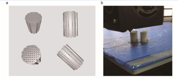
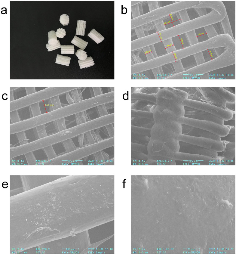
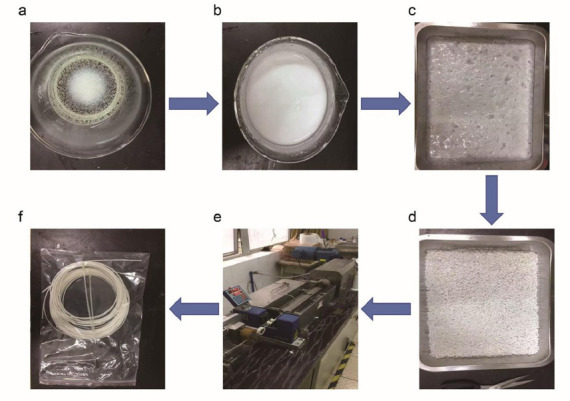
Methods: A bionic, porous HA/PCL scaffold was fabricated using 3D printing, and its microstructure, porosity, hydrophilicity, and mechanical properties were evaluated through scanning electron microscopy and various assays. Bone marrow mesenchymal stem cells (BMSCs) and vascular endothelial progenitor cells (VEPCs) were co-cultured with the scaffold, and their proliferation and osteogenic differentiation were assessed using the Cell Counting Kit-8, ALP assays, and alizarin red staining. Osteogenic marker expression was analyzed via qRT-PCR. In vivo bone regeneration was evaluated through histological analysis of H&E and Masson's trichrome staining in a rat cranial defect model.
Results: The average pore size of the scaffold was 462.00 ± 100.389 μm, with a porosity of 53%, a water absorption expansion rate of 5.10%, a contact angle of 94.55°, an elastic modulus of 53.82 MPa, and a compressive strength of 6.10 MPa. ALP activity and qRT-PCR analysis of osteogenic markers (BMP2, OCN, Runx2) showed significant upregulation in cells co-cultured with the scaffolds. In vivo experiments demonstrated enhanced bone regeneration and collagen deposition in the HA/PCL scaffold group.
Conclusion: The results suggest that the HA/PCL scaffold promotes osteogenic differentiation and bone regeneration, making it suitable for bone tissue engineering applications.
期刊介绍:
The Journal of Musculoskeletal and Neuronal Interactions (JMNI) is an academic journal dealing with the pathophysiology and treatment of musculoskeletal disorders. It is published quarterly (months of issue March, June, September, December). Its purpose is to publish original, peer-reviewed papers of research and clinical experience in all areas of the musculoskeletal system and its interactions with the nervous system, especially metabolic bone diseases, with particular emphasis on osteoporosis. Additionally, JMNI publishes the Abstracts from the biannual meetings of the International Society of Musculoskeletal and Neuronal Interactions, and hosts Abstracts of other meetings on topics related to the aims and scope of JMNI.
 求助内容:
求助内容: 应助结果提醒方式:


