Christina Schnoz, Marco Bonani, Florian Alexander Huber, Birgit Maria Helmchen, Thomas Fehr, Beata Bode-Lesniewska, Chantal Pauli, Ariana Gaspert
{"title":"Perinephric myxoid pseudotumor of fat - histopathological and molecular characterization of 3 cases after renal transplantation.","authors":"Christina Schnoz, Marco Bonani, Florian Alexander Huber, Birgit Maria Helmchen, Thomas Fehr, Beata Bode-Lesniewska, Chantal Pauli, Ariana Gaspert","doi":"10.1186/s13000-025-01615-4","DOIUrl":null,"url":null,"abstract":"<p><strong>Background: </strong>Perinephric myxoid pseudotumor of fat (PMPF) is a rare benign pseudo-neoplastic proliferation of the perinephric and renal sinus adipose tissue. Its pathogenesis is thought to be a reactive process typically associated with neoplastic and non-neoplastic end-stage kidney disease. The distinctive histopathological feature of PMPF is a myxoid process comprising bland, spindled stromal cells interspersed with mature adipose tissue. Macroscopically, it is characterized by tumorous lipomatous remodeling of the kidney, which may raise concerns of malignancy on imaging. To date, only seven cases of PMPF have been documented in the context of kidney transplantation.</p><p><strong>Case presentation: </strong>This report describes three cases of PMPF in patients following renal transplantation, involving both native and grafted kidneys. Macroscopically, all cases consisted of shrunken kidneys with thinned and atrophic renal parenchyma surrounded by massively hypertrophic perirenal fat with mass-forming nodules, which was in concordance with cross sectional imaging findings acquired before surgery. Histology of the remaining renal parenchyma showed end stage renal disease in all four surgically removed kidneys, with diffuse interstitial fibrosis, tubular atrophy and sclerosed glomeruli. Perirenal adipose tissue consisted of mature fat with areas of significant myxoid and collagenous stromal component, interspersed with bland spindle and stellate-shaped cells. Immunohistochemistry for S100, smooth muscle actin, desmin and IgG4 were negative. No MDM2 gene amplification was identified by fluorescence in situ hybridization. Broad molecular profiling using the FoundationOne<sup>®</sup>Heme assay revealed no evidence of pathogenic alterations on DNA and RNA levels.</p><p><strong>Conclusion: </strong>PMPF is a rare benign condition typically associated with chronic kidney disease, occurring late in the course. The radiological findings may be mistaken for those of a malignant tumor, and histopathological examination is required to exclude a malignant neoplasm, in particular a well-differentiated or dedifferentiated liposarcoma of the retroperitoneum. Renal transplant recipients can be affected by PMPF, which can occur in both native and transplanted kidneys several years following renal transplantation.</p>","PeriodicalId":11237,"journal":{"name":"Diagnostic Pathology","volume":"20 1","pages":"25"},"PeriodicalIF":2.3000,"publicationDate":"2025-03-01","publicationTypes":"Journal Article","fieldsOfStudy":null,"isOpenAccess":false,"openAccessPdf":"https://www.ncbi.nlm.nih.gov/pmc/articles/PMC11872312/pdf/","citationCount":"0","resultStr":null,"platform":"Semanticscholar","paperid":null,"PeriodicalName":"Diagnostic Pathology","FirstCategoryId":"3","ListUrlMain":"https://doi.org/10.1186/s13000-025-01615-4","RegionNum":3,"RegionCategory":"医学","ArticlePicture":[],"TitleCN":null,"AbstractTextCN":null,"PMCID":null,"EPubDate":"","PubModel":"","JCR":"Q2","JCRName":"PATHOLOGY","Score":null,"Total":0}
引用次数: 0
Abstract
Background: Perinephric myxoid pseudotumor of fat (PMPF) is a rare benign pseudo-neoplastic proliferation of the perinephric and renal sinus adipose tissue. Its pathogenesis is thought to be a reactive process typically associated with neoplastic and non-neoplastic end-stage kidney disease. The distinctive histopathological feature of PMPF is a myxoid process comprising bland, spindled stromal cells interspersed with mature adipose tissue. Macroscopically, it is characterized by tumorous lipomatous remodeling of the kidney, which may raise concerns of malignancy on imaging. To date, only seven cases of PMPF have been documented in the context of kidney transplantation.
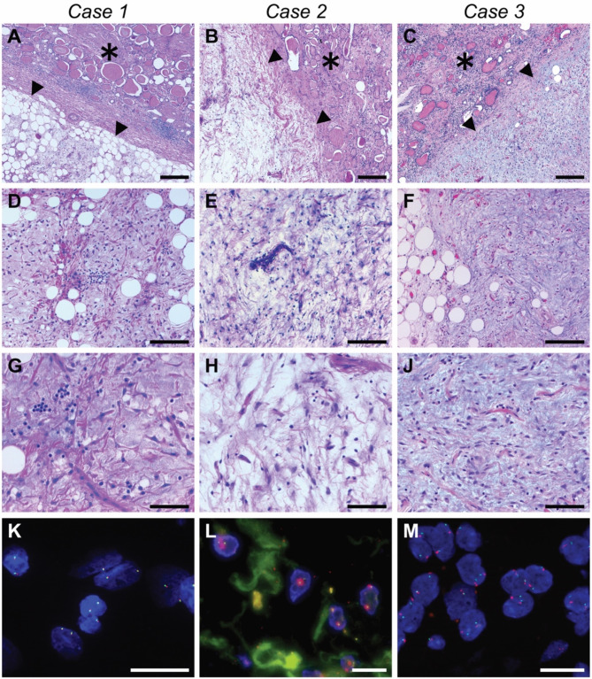
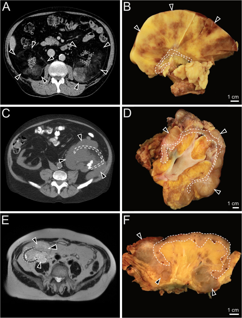
Case presentation: This report describes three cases of PMPF in patients following renal transplantation, involving both native and grafted kidneys. Macroscopically, all cases consisted of shrunken kidneys with thinned and atrophic renal parenchyma surrounded by massively hypertrophic perirenal fat with mass-forming nodules, which was in concordance with cross sectional imaging findings acquired before surgery. Histology of the remaining renal parenchyma showed end stage renal disease in all four surgically removed kidneys, with diffuse interstitial fibrosis, tubular atrophy and sclerosed glomeruli. Perirenal adipose tissue consisted of mature fat with areas of significant myxoid and collagenous stromal component, interspersed with bland spindle and stellate-shaped cells. Immunohistochemistry for S100, smooth muscle actin, desmin and IgG4 were negative. No MDM2 gene amplification was identified by fluorescence in situ hybridization. Broad molecular profiling using the FoundationOne®Heme assay revealed no evidence of pathogenic alterations on DNA and RNA levels.
Conclusion: PMPF is a rare benign condition typically associated with chronic kidney disease, occurring late in the course. The radiological findings may be mistaken for those of a malignant tumor, and histopathological examination is required to exclude a malignant neoplasm, in particular a well-differentiated or dedifferentiated liposarcoma of the retroperitoneum. Renal transplant recipients can be affected by PMPF, which can occur in both native and transplanted kidneys several years following renal transplantation.
期刊介绍:
Diagnostic Pathology is an open access, peer-reviewed, online journal that considers research in surgical and clinical pathology, immunology, and biology, with a special focus on cutting-edge approaches in diagnostic pathology and tissue-based therapy. The journal covers all aspects of surgical pathology, including classic diagnostic pathology, prognosis-related diagnosis (tumor stages, prognosis markers, such as MIB-percentage, hormone receptors, etc.), and therapy-related findings. The journal also focuses on the technological aspects of pathology, including molecular biology techniques, morphometry aspects (stereology, DNA analysis, syntactic structure analysis), communication aspects (telecommunication, virtual microscopy, virtual pathology institutions, etc.), and electronic education and quality assurance (for example interactive publication, on-line references with automated updating, etc.).

 求助内容:
求助内容: 应助结果提醒方式:


