Njål Lura, Kari S Wagner-Larsen, Stian Ryste, Kristine Fasmer, David Forsse, Jone Trovik, Mari K Halle, Bjørn I Bertelsen, Frank Riemer, Øyvind Salvesen, Kathrine Woie, Camilla Krakstad, Ingfrid S Haldorsen
{"title":"Tumor ADC value predicts outcome and yields refined prognostication in uterine cervical cancer.","authors":"Njål Lura, Kari S Wagner-Larsen, Stian Ryste, Kristine Fasmer, David Forsse, Jone Trovik, Mari K Halle, Bjørn I Bertelsen, Frank Riemer, Øyvind Salvesen, Kathrine Woie, Camilla Krakstad, Ingfrid S Haldorsen","doi":"10.1186/s40644-025-00828-6","DOIUrl":null,"url":null,"abstract":"<p><p>Pelvic MRI is essential for evaluating local and regional tumor extent in uterine cervical cancer (CC). Tumor microstructure captured by diffusion-weighted imaging (DWI) and apparent diffusion coefficient (ADC) markers may be closely linked to prognosis in CC.Purpose To explore whether primary tumor ADC markers predict survival in CC.Material and methods CC patients (n = 179) diagnosed during 2009-2020 with MRI-assessed primary maximum tumor<sub>size</sub> ≥ 2 cm were included in this retrospective single-center study. Two radiologists read all MRIs independently, measuring mean tumor ADC values in manually drawn regions of interest (ROIs) and mean tumor ADC (tumor<sub>ADCmean</sub>) from five measurements for the two readers was used. ADC from ROIs in the myometrium (myometrium<sub>ADC</sub>), cervical stroma (cervix<sub>ADC</sub>), and bladder (bladder<sub>ADC</sub>) were used to calculate ADC ratios. ADC markers were explored in relation to the International Federation of Gynecology and Obstetrics (FIGO) (2018) stage, disease-specific survival (DSS), and recurrence/progression-free survival (RPFS).Results Inter-reader agreement for all ADC measurements was high (ICC:0.59-0.79). Low tumor<sub>ADCmean</sub> predicted advanced FIGO stage (P = 0.04) and reduced DSS (hazard ratio (HR): 0.96, P < 0.001; AIC: 441). Myometrium<sub>ADC</sub>/tumor<sub>ADCmean</sub> yielded the best Cox regression fit (AIC = 430) among all tumor ADC markers. Patients with high myometrium<sub>ADC</sub>/tumor<sub>ADCmean</sub> had significantly reduced 5-year DSS for FIGO stage I, II, and III (P = 0.01, 0.004, and 0.02, respectively) and tended to the same for FIGO IV (P = 0.22).Conclusion Low tumor<sub>ADCmean</sub> predicted reduced DSS in CC. High myometrium<sub>ADC</sub>/tumor<sub>ADCmean</sub> was the strongest ADC predictor of poor DSS and a marker of high-risk phenotype independent of FIGO stage.</p>","PeriodicalId":9548,"journal":{"name":"Cancer Imaging","volume":"25 1","pages":"23"},"PeriodicalIF":3.5000,"publicationDate":"2025-02-28","publicationTypes":"Journal Article","fieldsOfStudy":null,"isOpenAccess":false,"openAccessPdf":"https://www.ncbi.nlm.nih.gov/pmc/articles/PMC11871606/pdf/","citationCount":"0","resultStr":null,"platform":"Semanticscholar","paperid":null,"PeriodicalName":"Cancer Imaging","FirstCategoryId":"3","ListUrlMain":"https://doi.org/10.1186/s40644-025-00828-6","RegionNum":2,"RegionCategory":"医学","ArticlePicture":[],"TitleCN":null,"AbstractTextCN":null,"PMCID":null,"EPubDate":"","PubModel":"","JCR":"Q2","JCRName":"ONCOLOGY","Score":null,"Total":0}
引用次数: 0
Abstract
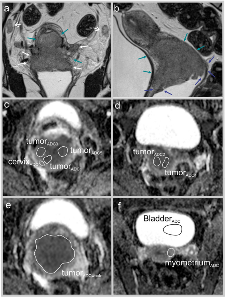
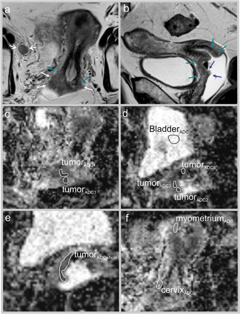
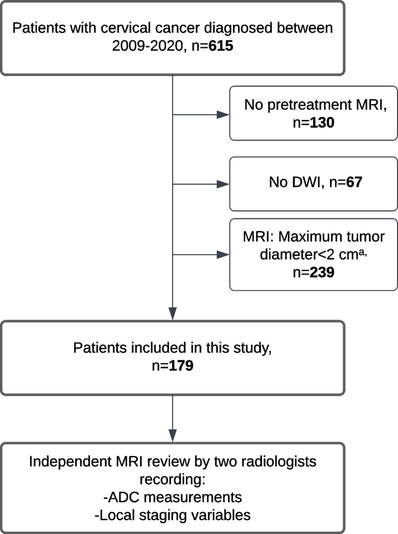
Pelvic MRI is essential for evaluating local and regional tumor extent in uterine cervical cancer (CC). Tumor microstructure captured by diffusion-weighted imaging (DWI) and apparent diffusion coefficient (ADC) markers may be closely linked to prognosis in CC.Purpose To explore whether primary tumor ADC markers predict survival in CC.Material and methods CC patients (n = 179) diagnosed during 2009-2020 with MRI-assessed primary maximum tumorsize ≥ 2 cm were included in this retrospective single-center study. Two radiologists read all MRIs independently, measuring mean tumor ADC values in manually drawn regions of interest (ROIs) and mean tumor ADC (tumorADCmean) from five measurements for the two readers was used. ADC from ROIs in the myometrium (myometriumADC), cervical stroma (cervixADC), and bladder (bladderADC) were used to calculate ADC ratios. ADC markers were explored in relation to the International Federation of Gynecology and Obstetrics (FIGO) (2018) stage, disease-specific survival (DSS), and recurrence/progression-free survival (RPFS).Results Inter-reader agreement for all ADC measurements was high (ICC:0.59-0.79). Low tumorADCmean predicted advanced FIGO stage (P = 0.04) and reduced DSS (hazard ratio (HR): 0.96, P < 0.001; AIC: 441). MyometriumADC/tumorADCmean yielded the best Cox regression fit (AIC = 430) among all tumor ADC markers. Patients with high myometriumADC/tumorADCmean had significantly reduced 5-year DSS for FIGO stage I, II, and III (P = 0.01, 0.004, and 0.02, respectively) and tended to the same for FIGO IV (P = 0.22).Conclusion Low tumorADCmean predicted reduced DSS in CC. High myometriumADC/tumorADCmean was the strongest ADC predictor of poor DSS and a marker of high-risk phenotype independent of FIGO stage.
Cancer ImagingONCOLOGY-RADIOLOGY, NUCLEAR MEDICINE & MEDICAL IMAGING
CiteScore
7.00
自引率
0.00%
发文量
66
审稿时长
>12 weeks
期刊介绍:
Cancer Imaging is an open access, peer-reviewed journal publishing original articles, reviews and editorials written by expert international radiologists working in oncology.
The journal encompasses CT, MR, PET, ultrasound, radionuclide and multimodal imaging in all kinds of malignant tumours, plus new developments, techniques and innovations. Topics of interest include:
Breast Imaging
Chest
Complications of treatment
Ear, Nose & Throat
Gastrointestinal
Hepatobiliary & Pancreatic
Imaging biomarkers
Interventional
Lymphoma
Measurement of tumour response
Molecular functional imaging
Musculoskeletal
Neuro oncology
Nuclear Medicine
Paediatric.

 求助内容:
求助内容: 应助结果提醒方式:


