{"title":"Preoperative diagnosis of meningioma sinus invasion based on MRI radiomics and deep learning: a multicenter study.","authors":"Yuan Gui, Wei Hu, Jialiang Ren, Fuqiang Tang, Limei Wang, Fang Zhang, Jing Zhang","doi":"10.1186/s40644-025-00845-5","DOIUrl":null,"url":null,"abstract":"<p><strong>Objective: </strong>Exploring the construction of a fusion model that combines radiomics and deep learning (DL) features is of great significance for the precise preoperative diagnosis of meningioma sinus invasion.</p><p><strong>Materials and methods: </strong>This study retrospectively collected data from 601 patients with meningioma confirmed by surgical pathology. For each patient, 3948 radiomics features, 12,288 VGG features, 6144 ResNet features, and 3072 DenseNet features were extracted from MRI images. Thus, univariate logistic regression, correlation analysis, and the Boruta algorithm were applied for further feature dimension reduction, selecting radiomics and DL features highly associated with meningioma sinus invasion. Finally, diagnosis models were constructed using the random forest (RF) algorithm. Additionally, the diagnostic performance of different models was evaluated using receiver operating characteristic (ROC) curves, and AUC values of different models were compared using the DeLong test.</p><p><strong>Results: </strong>Ultimately, 21 features highly associated with meningioma sinus invasion were selected, including 6 radiomics features, 2 VGG features, 7 ResNet features, and 6 DenseNet features. Based on these features, five models were constructed: the radiomics model, VGG model, ResNet model, DenseNet model, and DL-radiomics (DLR) fusion model. This fusion model demonstrated superior diagnostic performance, with AUC values of 0.818, 0.814, and 0.769 in the training set, internal validation set, and independent external validation set, respectively. Furthermore, the results of the DeLong test indicated that there were significant differences between the fusion model and both the radiomics model and the VGG model (p < 0.05).</p><p><strong>Conclusions: </strong>The fusion model combining radiomics and DL features exhibits superior diagnostic performance in preoperative diagnosis of meningioma sinus invasion. It is expected to become a powerful tool for clinical surgical plan selection and patient prognosis assessment.</p>","PeriodicalId":9548,"journal":{"name":"Cancer Imaging","volume":"25 1","pages":"20"},"PeriodicalIF":3.5000,"publicationDate":"2025-02-28","publicationTypes":"Journal Article","fieldsOfStudy":null,"isOpenAccess":false,"openAccessPdf":"https://www.ncbi.nlm.nih.gov/pmc/articles/PMC11869444/pdf/","citationCount":"0","resultStr":null,"platform":"Semanticscholar","paperid":null,"PeriodicalName":"Cancer Imaging","FirstCategoryId":"3","ListUrlMain":"https://doi.org/10.1186/s40644-025-00845-5","RegionNum":2,"RegionCategory":"医学","ArticlePicture":[],"TitleCN":null,"AbstractTextCN":null,"PMCID":null,"EPubDate":"","PubModel":"","JCR":"Q2","JCRName":"ONCOLOGY","Score":null,"Total":0}
引用次数: 0
Abstract
Objective: Exploring the construction of a fusion model that combines radiomics and deep learning (DL) features is of great significance for the precise preoperative diagnosis of meningioma sinus invasion.
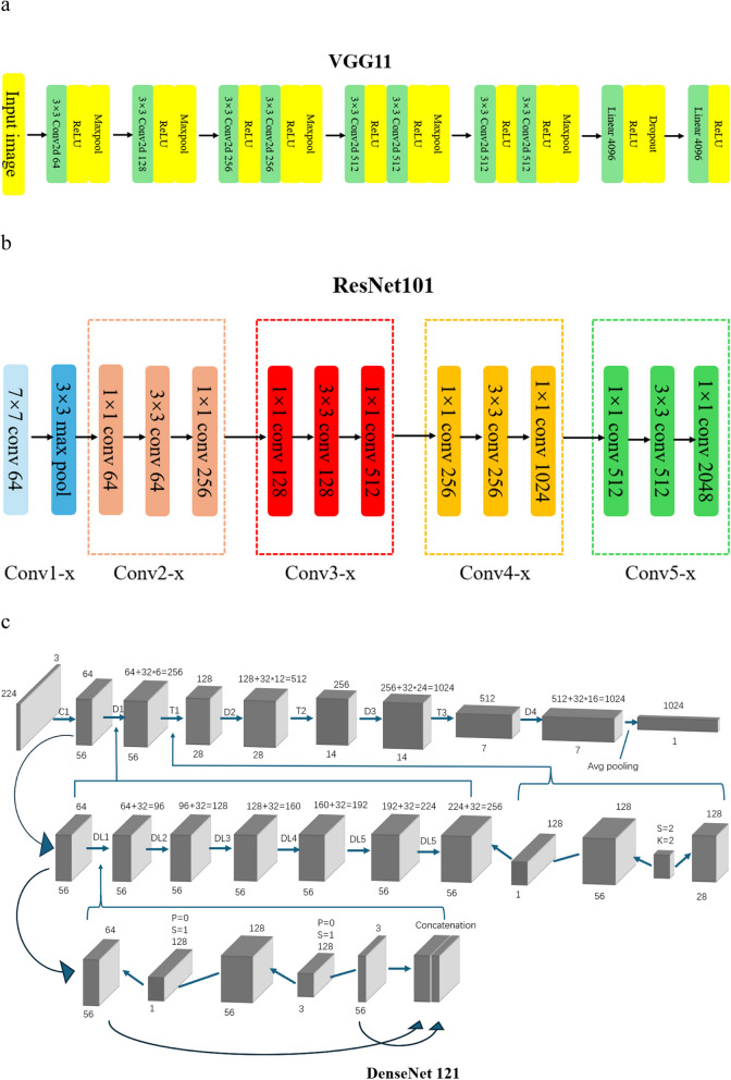
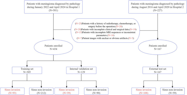
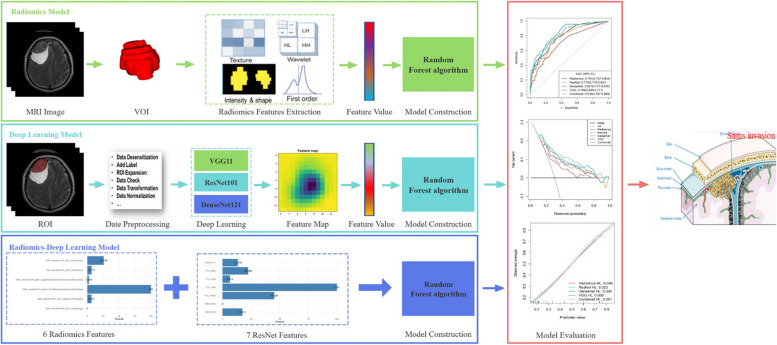
Materials and methods: This study retrospectively collected data from 601 patients with meningioma confirmed by surgical pathology. For each patient, 3948 radiomics features, 12,288 VGG features, 6144 ResNet features, and 3072 DenseNet features were extracted from MRI images. Thus, univariate logistic regression, correlation analysis, and the Boruta algorithm were applied for further feature dimension reduction, selecting radiomics and DL features highly associated with meningioma sinus invasion. Finally, diagnosis models were constructed using the random forest (RF) algorithm. Additionally, the diagnostic performance of different models was evaluated using receiver operating characteristic (ROC) curves, and AUC values of different models were compared using the DeLong test.
Results: Ultimately, 21 features highly associated with meningioma sinus invasion were selected, including 6 radiomics features, 2 VGG features, 7 ResNet features, and 6 DenseNet features. Based on these features, five models were constructed: the radiomics model, VGG model, ResNet model, DenseNet model, and DL-radiomics (DLR) fusion model. This fusion model demonstrated superior diagnostic performance, with AUC values of 0.818, 0.814, and 0.769 in the training set, internal validation set, and independent external validation set, respectively. Furthermore, the results of the DeLong test indicated that there were significant differences between the fusion model and both the radiomics model and the VGG model (p < 0.05).
Conclusions: The fusion model combining radiomics and DL features exhibits superior diagnostic performance in preoperative diagnosis of meningioma sinus invasion. It is expected to become a powerful tool for clinical surgical plan selection and patient prognosis assessment.
Cancer ImagingONCOLOGY-RADIOLOGY, NUCLEAR MEDICINE & MEDICAL IMAGING
CiteScore
7.00
自引率
0.00%
发文量
66
审稿时长
>12 weeks
期刊介绍:
Cancer Imaging is an open access, peer-reviewed journal publishing original articles, reviews and editorials written by expert international radiologists working in oncology.
The journal encompasses CT, MR, PET, ultrasound, radionuclide and multimodal imaging in all kinds of malignant tumours, plus new developments, techniques and innovations. Topics of interest include:
Breast Imaging
Chest
Complications of treatment
Ear, Nose & Throat
Gastrointestinal
Hepatobiliary & Pancreatic
Imaging biomarkers
Interventional
Lymphoma
Measurement of tumour response
Molecular functional imaging
Musculoskeletal
Neuro oncology
Nuclear Medicine
Paediatric.

 求助内容:
求助内容: 应助结果提醒方式:


