Fiona Dierksen, Johanna S Geibel, Janika Albrecht, Sabine Hofer, Peter Dechent, Amelie C Hesse, Jens Frahm, Mathias Bähr, Jan C Koch, Jan Liman, Ilko L Maier
{"title":"T1-relaxation times along the corticospinal tract as a diagnostic marker in patients with amyotrophic lateral sclerosis.","authors":"Fiona Dierksen, Johanna S Geibel, Janika Albrecht, Sabine Hofer, Peter Dechent, Amelie C Hesse, Jens Frahm, Mathias Bähr, Jan C Koch, Jan Liman, Ilko L Maier","doi":"10.3389/fnimg.2025.1549727","DOIUrl":null,"url":null,"abstract":"<p><strong>Background and purpose: </strong>In the differential diagnostic workup of amyotrophic lateral sclerosis (ALS), magnetic resonance imaging (MRI) is primarily used to rule out significant differential diagnoses. So far, whole-brain T1-mapping has not been assessed as a diagnostic tool in this patient population.</p><p><strong>Methods: </strong>We investigated the diagnostic potential of a novel T1-mapping method based on real-time MRI with 0.5 mm in-plane resolution and 4s acquisition time per slice. The study included patients aged 18 to 90 years who met the revised El Escorial criteria for at least possible ALS. T1-relaxation times were measured along the corticospinal tract in predefined regions of interest.</p><p><strong>Results: </strong>Twenty-nine ALS-patients and 43 control group patients (CG) were included in the study. Median ALS Functional Rating Scale revised (ALSFRS-R) was 37 (IQR, 35-44) points and the mean duration from symptom onset to MRI was 21 ± 17 (SD) months. ALS patients showed significantly higher T1-relaxation times in all ROIs compared to CG with mean differences in the hand knob of 50 ms (<i>p</i> < 0.001), corona radiata 24 ms (<i>p</i> = 0.034), internal capsule 27 ms (<i>p</i> = 0.002) and midbrain peduncles 48 ms (<i>p</i> < 0.001). There was a consistent negative correlation between the ALSFRS-R and T1-relaxation times in all ROIs.</p><p><strong>Conclusions: </strong>T1-relaxation times along the corticospinal tract are significantly elevated in ALS patients compared to CG and associated with lower ALSFRS-R. These results imply the analysis of T1-relaxation times as a promising diagnostic tool that can distinguish ALS patients from the control group. Ongoing longitudinal studies may provide deeper insights into disease progression and the effects of therapeutic interventions.</p>","PeriodicalId":73094,"journal":{"name":"Frontiers in neuroimaging","volume":"4 ","pages":"1549727"},"PeriodicalIF":0.0000,"publicationDate":"2025-02-13","publicationTypes":"Journal Article","fieldsOfStudy":null,"isOpenAccess":false,"openAccessPdf":"https://www.ncbi.nlm.nih.gov/pmc/articles/PMC11865248/pdf/","citationCount":"0","resultStr":null,"platform":"Semanticscholar","paperid":null,"PeriodicalName":"Frontiers in neuroimaging","FirstCategoryId":"1085","ListUrlMain":"https://doi.org/10.3389/fnimg.2025.1549727","RegionNum":0,"RegionCategory":null,"ArticlePicture":[],"TitleCN":null,"AbstractTextCN":null,"PMCID":null,"EPubDate":"2025/1/1 0:00:00","PubModel":"eCollection","JCR":"","JCRName":"","Score":null,"Total":0}
引用次数: 0
Abstract
Background and purpose: In the differential diagnostic workup of amyotrophic lateral sclerosis (ALS), magnetic resonance imaging (MRI) is primarily used to rule out significant differential diagnoses. So far, whole-brain T1-mapping has not been assessed as a diagnostic tool in this patient population.
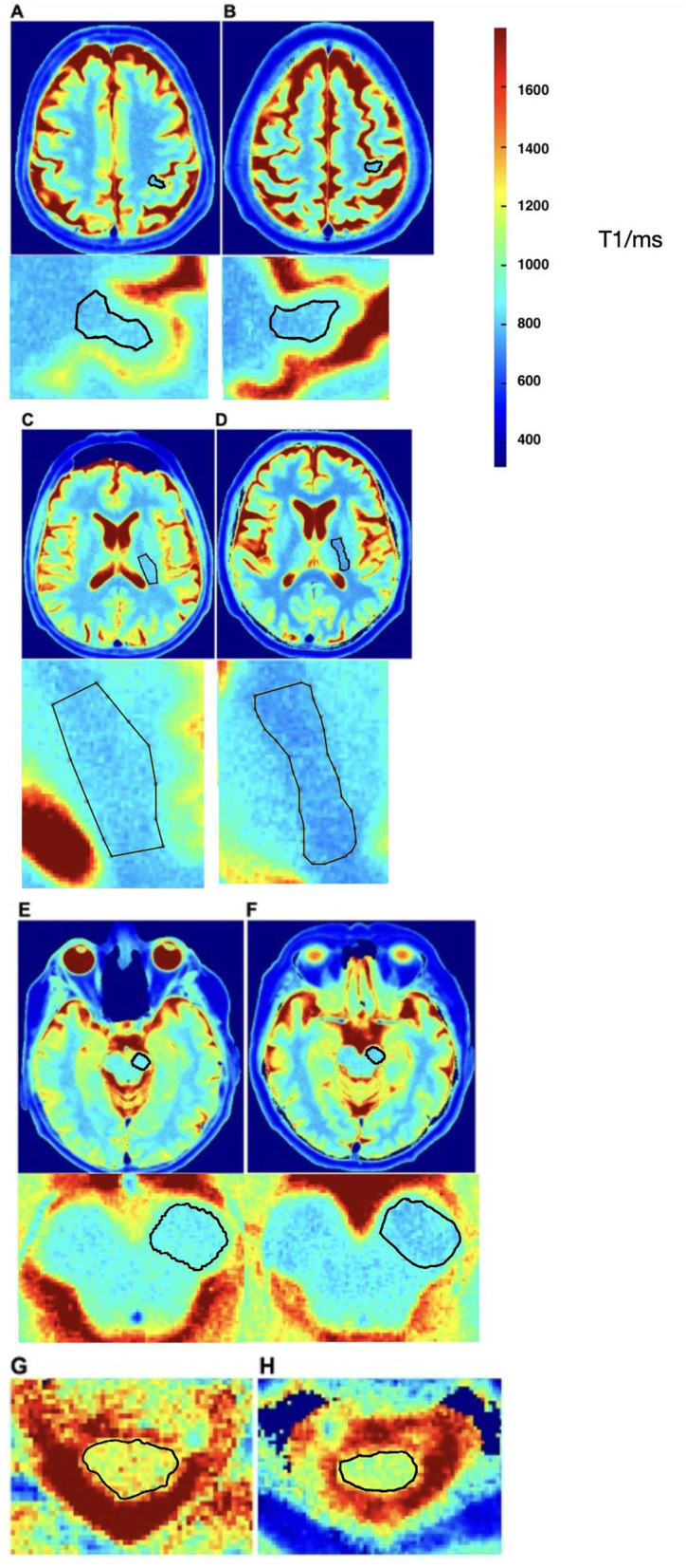
Methods: We investigated the diagnostic potential of a novel T1-mapping method based on real-time MRI with 0.5 mm in-plane resolution and 4s acquisition time per slice. The study included patients aged 18 to 90 years who met the revised El Escorial criteria for at least possible ALS. T1-relaxation times were measured along the corticospinal tract in predefined regions of interest.
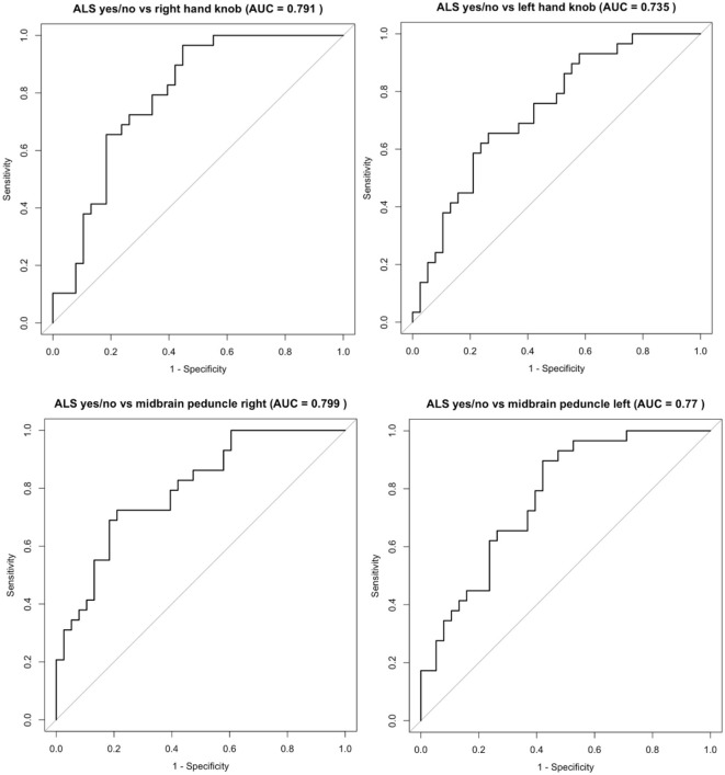
Results: Twenty-nine ALS-patients and 43 control group patients (CG) were included in the study. Median ALS Functional Rating Scale revised (ALSFRS-R) was 37 (IQR, 35-44) points and the mean duration from symptom onset to MRI was 21 ± 17 (SD) months. ALS patients showed significantly higher T1-relaxation times in all ROIs compared to CG with mean differences in the hand knob of 50 ms (p < 0.001), corona radiata 24 ms (p = 0.034), internal capsule 27 ms (p = 0.002) and midbrain peduncles 48 ms (p < 0.001). There was a consistent negative correlation between the ALSFRS-R and T1-relaxation times in all ROIs.
Conclusions: T1-relaxation times along the corticospinal tract are significantly elevated in ALS patients compared to CG and associated with lower ALSFRS-R. These results imply the analysis of T1-relaxation times as a promising diagnostic tool that can distinguish ALS patients from the control group. Ongoing longitudinal studies may provide deeper insights into disease progression and the effects of therapeutic interventions.

 求助内容:
求助内容: 应助结果提醒方式:


