Combination of CT/MRI LI-RADS With Second-Line Contrast-Enhanced Ultrasound Using Sulfur Hexafluoride or Perfluorobutane for Diagnosing Hepatocellular Carcinoma in High-Risk Patients.
IF 5.3 2区 医学Q1 RADIOLOGY, NUCLEAR MEDICINE & MEDICAL IMAGING
Yu Li, Sheng Li, Qing Li, Kai Li, Jing Han, Siyue Mao, Xiaohong Xu, Zhongzhen Su, Yanling Zuo, Shousong Xie, Hong Wen, Xuebin Zou, Jingxian Shen, Lingling Li, Jianhua Zhou
{"title":"Combination of CT/MRI LI-RADS With Second-Line Contrast-Enhanced Ultrasound Using Sulfur Hexafluoride or Perfluorobutane for Diagnosing Hepatocellular Carcinoma in High-Risk Patients.","authors":"Yu Li, Sheng Li, Qing Li, Kai Li, Jing Han, Siyue Mao, Xiaohong Xu, Zhongzhen Su, Yanling Zuo, Shousong Xie, Hong Wen, Xuebin Zou, Jingxian Shen, Lingling Li, Jianhua Zhou","doi":"10.3348/kjr.2024.0980","DOIUrl":null,"url":null,"abstract":"<p><strong>Objective: </strong>The CT/MRI Liver Imaging Reporting and Data System (LI-RADS) demonstrates high specificity with relatively limited sensitivity for diagnosing hepatocellular carcinoma (HCC) in high-risk patients. This study aimed to explore the possibility of improving sensitivity by combining CT/MRI LI-RADS v2018 with second-line contrast-enhanced ultrasound (CEUS) LI-RADS v2017 using sulfur hexafluoride (SHF) or perfluorobutane (PFB).</p><p><strong>Materials and methods: </strong>This retrospective analysis of prospectively collected multicenter data included high-risk patients with treatment-naive hepatic observations. The reference standard was pathological confirmation or a composite reference standard (only for benign lesions). Each participant underwent concurrent CT/MRI, SHF-enhanced US, and PFB-enhanced US examinations. The diagnostic performances for HCC of CT/MRI LI-RADS alone and three combination strategies (combining CT/MRI LI-RADS with either LI-RADS SHF, LI-RADS PFB, or a modified algorithm incorporating the Kupffer-phase findings for PFB [modified PFB]) were evaluated. For the three combination strategies, apart from the CT/MRI LR-5 criteria, HCC was diagnosed if CT/MRI LR-3 or LR-4 observations met the LR-5 criteria using LI-RADS SHF, LI-RADS PFB, or modified PFB.</p><p><strong>Results: </strong>In total, 281 participants (237 males; mean age, 55 ± 11 years) with 306 observations (227 HCCs, 40 non-HCC malignancies, and 39 benign lesions) were included. Using LI-RADS SHF, LI-RADS PFB, and modified PFB, 20, 23, and 31 CT/MRI LR-3/4 observations, respectively, were reclassified as LR-5, and all were pathologically confirmed as HCCs. Compared to CT/MRI LI-RADS alone (74%, 95% confidence interval [CI]: 68%-79%), the three combination strategies combining CT/MRI LI-RADS with either LI-RADS SHF, LI-RADS PFB, or modified PFB increased sensitivity (83% [95% CI: 77%-87%], 84% [95% CI: 79%-89%], 88% [95% CI: 83%-92%], respectively; all <i>P</i> < 0.001), while maintaining the specificity at 92% (95% CI: 84%-97%).</p><p><strong>Conclusion: </strong>The combination of CT/MRI LI-RADS with second-line CEUS using SHF or PFB improved the sensitivity of HCC diagnosis without compromising specificity.</p>","PeriodicalId":17881,"journal":{"name":"Korean Journal of Radiology","volume":" ","pages":"346-359"},"PeriodicalIF":5.3000,"publicationDate":"2025-04-01","publicationTypes":"Journal Article","fieldsOfStudy":null,"isOpenAccess":false,"openAccessPdf":"https://www.ncbi.nlm.nih.gov/pmc/articles/PMC11955382/pdf/","citationCount":"0","resultStr":null,"platform":"Semanticscholar","paperid":null,"PeriodicalName":"Korean Journal of Radiology","FirstCategoryId":"3","ListUrlMain":"https://doi.org/10.3348/kjr.2024.0980","RegionNum":2,"RegionCategory":"医学","ArticlePicture":[],"TitleCN":null,"AbstractTextCN":null,"PMCID":null,"EPubDate":"2025/2/20 0:00:00","PubModel":"Epub","JCR":"Q1","JCRName":"RADIOLOGY, NUCLEAR MEDICINE & MEDICAL IMAGING","Score":null,"Total":0}
引用次数: 0
Abstract
Objective: The CT/MRI Liver Imaging Reporting and Data System (LI-RADS) demonstrates high specificity with relatively limited sensitivity for diagnosing hepatocellular carcinoma (HCC) in high-risk patients. This study aimed to explore the possibility of improving sensitivity by combining CT/MRI LI-RADS v2018 with second-line contrast-enhanced ultrasound (CEUS) LI-RADS v2017 using sulfur hexafluoride (SHF) or perfluorobutane (PFB).
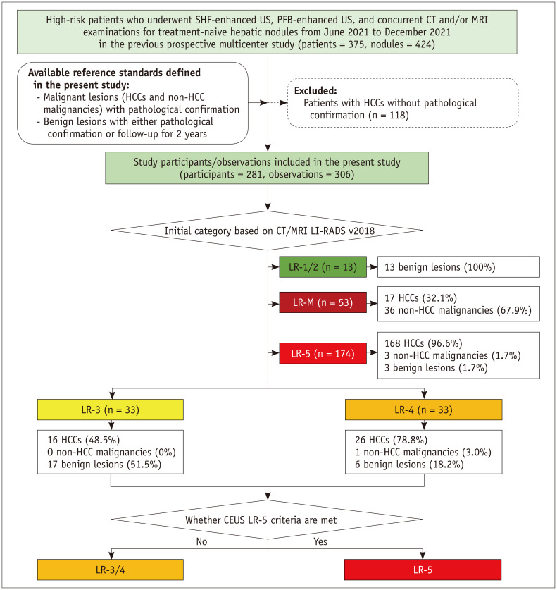
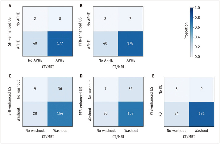
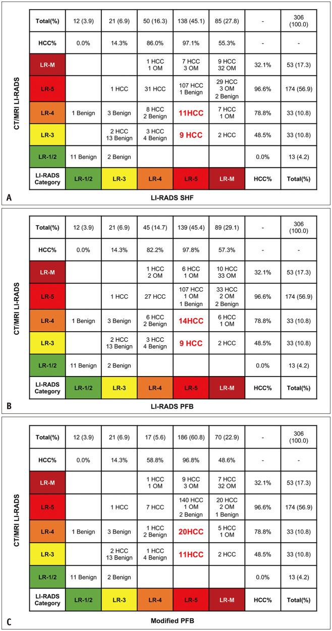
Materials and methods: This retrospective analysis of prospectively collected multicenter data included high-risk patients with treatment-naive hepatic observations. The reference standard was pathological confirmation or a composite reference standard (only for benign lesions). Each participant underwent concurrent CT/MRI, SHF-enhanced US, and PFB-enhanced US examinations. The diagnostic performances for HCC of CT/MRI LI-RADS alone and three combination strategies (combining CT/MRI LI-RADS with either LI-RADS SHF, LI-RADS PFB, or a modified algorithm incorporating the Kupffer-phase findings for PFB [modified PFB]) were evaluated. For the three combination strategies, apart from the CT/MRI LR-5 criteria, HCC was diagnosed if CT/MRI LR-3 or LR-4 observations met the LR-5 criteria using LI-RADS SHF, LI-RADS PFB, or modified PFB.
Results: In total, 281 participants (237 males; mean age, 55 ± 11 years) with 306 observations (227 HCCs, 40 non-HCC malignancies, and 39 benign lesions) were included. Using LI-RADS SHF, LI-RADS PFB, and modified PFB, 20, 23, and 31 CT/MRI LR-3/4 observations, respectively, were reclassified as LR-5, and all were pathologically confirmed as HCCs. Compared to CT/MRI LI-RADS alone (74%, 95% confidence interval [CI]: 68%-79%), the three combination strategies combining CT/MRI LI-RADS with either LI-RADS SHF, LI-RADS PFB, or modified PFB increased sensitivity (83% [95% CI: 77%-87%], 84% [95% CI: 79%-89%], 88% [95% CI: 83%-92%], respectively; all P < 0.001), while maintaining the specificity at 92% (95% CI: 84%-97%).
Conclusion: The combination of CT/MRI LI-RADS with second-line CEUS using SHF or PFB improved the sensitivity of HCC diagnosis without compromising specificity.
期刊介绍:
The inaugural issue of the Korean J Radiol came out in March 2000. Our journal aims to produce and propagate knowledge on radiologic imaging and related sciences.
A unique feature of the articles published in the Journal will be their reflection of global trends in radiology combined with an East-Asian perspective. Geographic differences in disease prevalence will be reflected in the contents of papers, and this will serve to enrich our body of knowledge.
World''s outstanding radiologists from many countries are serving as editorial board of our journal.

 求助内容:
求助内容: 应助结果提醒方式:


