{"title":"Influence of surface texture on osteogenic differentiation of dental pulp stem cells: An <i>in vitro</i> study.","authors":"Komal Rajpurohit, Vidya Dodwad, Avinash Kharat, Spoorthi Belludi, Pooja Pharne, Sarah Marium","doi":"10.4103/jisp.jisp_307_23","DOIUrl":null,"url":null,"abstract":"<p><strong>Background: </strong>In comparison with perfectly machined surface implants, surface topographic modifications like roughness accelerate the osteogenesis of dental pulpal stem cells (DPSC). This greatly enhances bone-implant contact and osteogenic potential of the stem cells. Hence, the aim of the current study was to evaluate and compare the differentiation and proliferation potential of stem cells obtained from dental pulp on sand-blasted and acid etched implant discs surfaces.</p><p><strong>Materials and methods: </strong>Stem cells from dental pulp were extracted from the premolar region of oral cavity. Titanium discs that measured one centimeter in diameter and three millimetres in thickness were used as investigation surfaces. Titanium surface disc were acid etched and sandblasted. Investigation had three group: acid etched (Group A), sandblasted (Group B), and standard control group, i.e., cells treated with osteogenic induction media only (Group C). In Group C, mesenchymal stem cells (MSCs) were treated with osteogenic induction medium without any titanium disc and these cells were used as standard controls. To identify which modified implant surface had greater potential for proliferation, 3-(4,5-dimethylthiazol-2-yl)-2,5-diphenyltetrazolium bromide (MTT) assay was performed using the explant culture. MTT assay assessed the viability of the cells as a function of its redox potential. This was followed by recognition of the stem cells for CD90, CD73, and CD 105 markers using flow cytometry with RUNX2 antibody on days 7 and 21 of incubation. The isolated cells were stained using 1% alizarin red stain to identify the number of stem cells per square centimeter area under the light microscope.</p><p><strong>Results: </strong>The osteogenic differentiation of both the materials was compared with standard control (MSCs treated with osteogenic differentiation media only). The osteoblastic cells on the acid-etched and sand-blasted implant surface disc had an almost identical capacity for proliferation till the MTT assay but according to the results of the alizarin red staining there was a slightly higher proliferation potential on acid etched surfaces compared to the sand blasted surfaces. Therefore, acid etched surfaces showed higher potential of osteogenic differentiation of DPSCs compared with sand-blasted surfaces.</p><p><strong>Conclusion: </strong>In comparison with perfectly machined surface implants, topographic surface modifications such as roughness can accelerate the osteogenesis of DPSC <i>in</i> <i>vitro</i>.</p>","PeriodicalId":15890,"journal":{"name":"Journal of Indian Society of Periodontology","volume":"28 4","pages":"478-483"},"PeriodicalIF":0.0000,"publicationDate":"2024-07-01","publicationTypes":"Journal Article","fieldsOfStudy":null,"isOpenAccess":false,"openAccessPdf":"https://www.ncbi.nlm.nih.gov/pmc/articles/PMC11864341/pdf/","citationCount":"0","resultStr":null,"platform":"Semanticscholar","paperid":null,"PeriodicalName":"Journal of Indian Society of Periodontology","FirstCategoryId":"1085","ListUrlMain":"https://doi.org/10.4103/jisp.jisp_307_23","RegionNum":0,"RegionCategory":null,"ArticlePicture":[],"TitleCN":null,"AbstractTextCN":null,"PMCID":null,"EPubDate":"2025/1/6 0:00:00","PubModel":"Epub","JCR":"Q2","JCRName":"Dentistry","Score":null,"Total":0}
引用次数: 0
Abstract
Background: In comparison with perfectly machined surface implants, surface topographic modifications like roughness accelerate the osteogenesis of dental pulpal stem cells (DPSC). This greatly enhances bone-implant contact and osteogenic potential of the stem cells. Hence, the aim of the current study was to evaluate and compare the differentiation and proliferation potential of stem cells obtained from dental pulp on sand-blasted and acid etched implant discs surfaces.
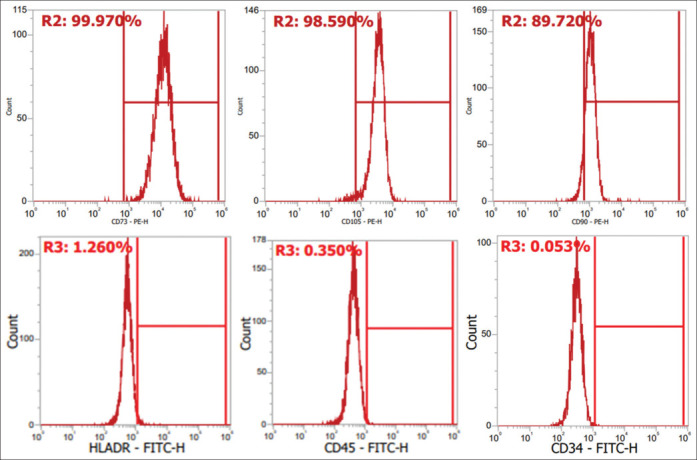
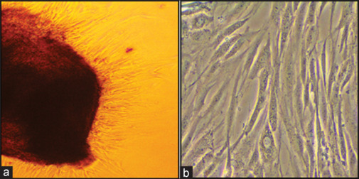
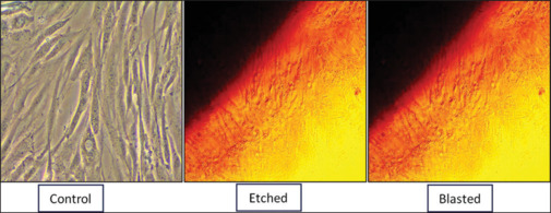
Materials and methods: Stem cells from dental pulp were extracted from the premolar region of oral cavity. Titanium discs that measured one centimeter in diameter and three millimetres in thickness were used as investigation surfaces. Titanium surface disc were acid etched and sandblasted. Investigation had three group: acid etched (Group A), sandblasted (Group B), and standard control group, i.e., cells treated with osteogenic induction media only (Group C). In Group C, mesenchymal stem cells (MSCs) were treated with osteogenic induction medium without any titanium disc and these cells were used as standard controls. To identify which modified implant surface had greater potential for proliferation, 3-(4,5-dimethylthiazol-2-yl)-2,5-diphenyltetrazolium bromide (MTT) assay was performed using the explant culture. MTT assay assessed the viability of the cells as a function of its redox potential. This was followed by recognition of the stem cells for CD90, CD73, and CD 105 markers using flow cytometry with RUNX2 antibody on days 7 and 21 of incubation. The isolated cells were stained using 1% alizarin red stain to identify the number of stem cells per square centimeter area under the light microscope.
Results: The osteogenic differentiation of both the materials was compared with standard control (MSCs treated with osteogenic differentiation media only). The osteoblastic cells on the acid-etched and sand-blasted implant surface disc had an almost identical capacity for proliferation till the MTT assay but according to the results of the alizarin red staining there was a slightly higher proliferation potential on acid etched surfaces compared to the sand blasted surfaces. Therefore, acid etched surfaces showed higher potential of osteogenic differentiation of DPSCs compared with sand-blasted surfaces.
Conclusion: In comparison with perfectly machined surface implants, topographic surface modifications such as roughness can accelerate the osteogenesis of DPSC invitro.
期刊介绍:
The Journal of Indian Society of Periodontology publishes original scientific articles to support practice , education and research in the dental specialty of periodontology and oral implantology. Journal of Indian Society of Periodontology (JISP), is the official publication of the Society and is managed and brought out by the Editor of the society. The journal is published Bimonthly with special issues being brought out for specific occasions. The ISP had a bulletin as its publication for a large number of years and was enhanced as a Journal a few years ago

 求助内容:
求助内容: 应助结果提醒方式:


