Lefkothea Pantazi, Valérie Untereiner, Paolo Rosales, Romain Rivet, Sandra Audonnet, Isabelle Proult, Laurent Ramont, Ganesh D. Sockalingum and Stéphane Brézillon
{"title":"Extracellular vesicles derived from ovarian cancer cell lines discriminated by biochemical and Fourier transform infrared spectroscopy approaches†","authors":"Lefkothea Pantazi, Valérie Untereiner, Paolo Rosales, Romain Rivet, Sandra Audonnet, Isabelle Proult, Laurent Ramont, Ganesh D. Sockalingum and Stéphane Brézillon","doi":"10.1039/D5AN00024F","DOIUrl":null,"url":null,"abstract":"<p >Ovarian cancer is the most lethal cancer among gynaecological malignancies. Due to the lack of early symptoms and screening tools, patients are diagnosed in advanced stages. Cancer invasion and metastasis through the extracellular matrix (ECM) are enhanced by tumour cell Extracellular Vesicles (EV). The aim of this study was to characterise the EVs derived from two ovarian cancer cell lines (ES2 and SKOV3) using biochemical and vibrational spectroscopic approaches. EVs were prepared by ultracentrifugation and characterised by Nanoparticle Tracking Analysis. Their surface proteins were assessed by MACSPlex EV kit for human exosomes. The presence of MMP14 and integrin subunits was evaluated in EVs and cell protein extracts by Western immunoblotting. Both EVs and cells were measured by Fourier transform infrared spectroscopy (FTIR) and data were analysed by hierarchical cluster analysis (HCA). Spectral differences were observed in the lipids and polysaccharides regions both between the SKOV3 and ES2 cells and their corresponding EVs, which allowed a good delineation by HCA. The differences in the biochemical data were confirmed by similar and specific features exhibited in their respective infrared spectral signatures. ES2 EVs exhibited an enrichment in MMP14 in agreement with the aggressiveness of this ovarian cancer metastatic cell line.</p>","PeriodicalId":63,"journal":{"name":"Analyst","volume":" 7","pages":" 1280-1292"},"PeriodicalIF":3.6000,"publicationDate":"2025-02-28","publicationTypes":"Journal Article","fieldsOfStudy":null,"isOpenAccess":false,"openAccessPdf":"","citationCount":"0","resultStr":null,"platform":"Semanticscholar","paperid":null,"PeriodicalName":"Analyst","FirstCategoryId":"92","ListUrlMain":"https://pubs.rsc.org/en/content/articlelanding/2025/an/d5an00024f","RegionNum":3,"RegionCategory":"化学","ArticlePicture":[],"TitleCN":null,"AbstractTextCN":null,"PMCID":null,"EPubDate":"","PubModel":"","JCR":"Q2","JCRName":"CHEMISTRY, ANALYTICAL","Score":null,"Total":0}
引用次数: 0
Abstract
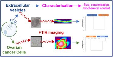
Ovarian cancer is the most lethal cancer among gynaecological malignancies. Due to the lack of early symptoms and screening tools, patients are diagnosed in advanced stages. Cancer invasion and metastasis through the extracellular matrix (ECM) are enhanced by tumour cell Extracellular Vesicles (EV). The aim of this study was to characterise the EVs derived from two ovarian cancer cell lines (ES2 and SKOV3) using biochemical and vibrational spectroscopic approaches. EVs were prepared by ultracentrifugation and characterised by Nanoparticle Tracking Analysis. Their surface proteins were assessed by MACSPlex EV kit for human exosomes. The presence of MMP14 and integrin subunits was evaluated in EVs and cell protein extracts by Western immunoblotting. Both EVs and cells were measured by Fourier transform infrared spectroscopy (FTIR) and data were analysed by hierarchical cluster analysis (HCA). Spectral differences were observed in the lipids and polysaccharides regions both between the SKOV3 and ES2 cells and their corresponding EVs, which allowed a good delineation by HCA. The differences in the biochemical data were confirmed by similar and specific features exhibited in their respective infrared spectral signatures. ES2 EVs exhibited an enrichment in MMP14 in agreement with the aggressiveness of this ovarian cancer metastatic cell line.

 求助内容:
求助内容: 应助结果提醒方式:


