{"title":"Bilateral blindness in a cat after a dental procedure, suspected to be due to segmental chorioretinal ischaemia necrosis.","authors":"Cleo Guerreiro, Christine Heinrich, Karen Walsh","doi":"10.1177/20551169251313619","DOIUrl":null,"url":null,"abstract":"<p><strong>Case summary: </strong>A 5-year-old female spayed domestic shorthair cat presented for sudden onset vision loss 3 days after a dental procedure. Bilateral blindness was confirmed on ocular examination, with fundoscopy revealing segmental wedge-shaped areas of retinal oedema and partial non-rhegmatogenous retinal detachments. An initial differential diagnosis included angioinvasive pulmonary carcinoma, based on previously reported fundoscopy images of this condition; however, general physical examination, blood pressure and chest radiographs were all normal. Four weeks after initial presentation, fundoscopy revealed the resolution of the retinal detachments; however, bilateral segmental chorioretinal necrosis was present. The cat regained some vision and remained well 13 months after the initial presentation. Considering the clinical findings, the onset of blindness after a lengthy dental procedure and improvement of vision over time, a diagnosis of pulmonary carcinoma was unlikely and instead a diagnosis of chorioretinal ischaemia secondary to maxillary artery blood flow restriction is proposed.</p><p><strong>Relevance and novel information: </strong>Maxillary artery blood flow restriction has been well documented with varying degrees of jaw opening in cats. Presumed central blindness as a result of this blood flow restriction has also been documented. However, to the authors' knowledge, vision loss due to retinal changes, documented by fundoscopic images and their progression over time following suspected chorioretinal ischaemia after a dental procedure, have not previously been reported. We propose that temporary occlusion of the maxillary artery can result in segmental chorioretinal necrosis and associated blindness in cats. This finding further supports the recommendation to minimise prolonged jaw opening during surgical procedures in cats.</p>","PeriodicalId":36588,"journal":{"name":"Journal of Feline Medicine and Surgery Open Reports","volume":"11 1","pages":"20551169251313619"},"PeriodicalIF":0.7000,"publicationDate":"2025-02-25","publicationTypes":"Journal Article","fieldsOfStudy":null,"isOpenAccess":false,"openAccessPdf":"https://www.ncbi.nlm.nih.gov/pmc/articles/PMC11863224/pdf/","citationCount":"0","resultStr":null,"platform":"Semanticscholar","paperid":null,"PeriodicalName":"Journal of Feline Medicine and Surgery Open Reports","FirstCategoryId":"1085","ListUrlMain":"https://doi.org/10.1177/20551169251313619","RegionNum":0,"RegionCategory":null,"ArticlePicture":[],"TitleCN":null,"AbstractTextCN":null,"PMCID":null,"EPubDate":"2025/1/1 0:00:00","PubModel":"eCollection","JCR":"Q3","JCRName":"VETERINARY SCIENCES","Score":null,"Total":0}
引用次数: 0
Abstract
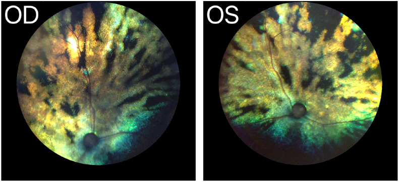
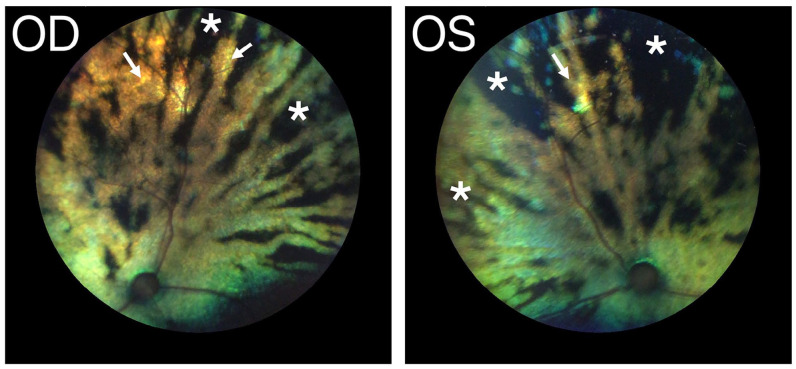
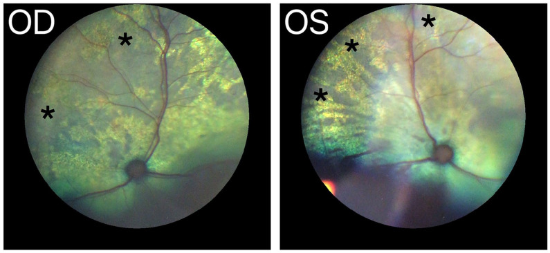
Case summary: A 5-year-old female spayed domestic shorthair cat presented for sudden onset vision loss 3 days after a dental procedure. Bilateral blindness was confirmed on ocular examination, with fundoscopy revealing segmental wedge-shaped areas of retinal oedema and partial non-rhegmatogenous retinal detachments. An initial differential diagnosis included angioinvasive pulmonary carcinoma, based on previously reported fundoscopy images of this condition; however, general physical examination, blood pressure and chest radiographs were all normal. Four weeks after initial presentation, fundoscopy revealed the resolution of the retinal detachments; however, bilateral segmental chorioretinal necrosis was present. The cat regained some vision and remained well 13 months after the initial presentation. Considering the clinical findings, the onset of blindness after a lengthy dental procedure and improvement of vision over time, a diagnosis of pulmonary carcinoma was unlikely and instead a diagnosis of chorioretinal ischaemia secondary to maxillary artery blood flow restriction is proposed.
Relevance and novel information: Maxillary artery blood flow restriction has been well documented with varying degrees of jaw opening in cats. Presumed central blindness as a result of this blood flow restriction has also been documented. However, to the authors' knowledge, vision loss due to retinal changes, documented by fundoscopic images and their progression over time following suspected chorioretinal ischaemia after a dental procedure, have not previously been reported. We propose that temporary occlusion of the maxillary artery can result in segmental chorioretinal necrosis and associated blindness in cats. This finding further supports the recommendation to minimise prolonged jaw opening during surgical procedures in cats.

 求助内容:
求助内容: 应助结果提醒方式:


