{"title":"Confronting Upside-Down Video-Assisted Thoracic Surgery Approach for Hemorrhagic Bronchogenic Cyst Manifested by Sudden Back Pain.","authors":"Masato Kambe, Tomonari Oki, Shuhei Iizuka, Yoshiro Otsuki, Toru Nakamura","doi":"10.70352/scrj.cr.24-0126","DOIUrl":null,"url":null,"abstract":"<p><strong>Introduction: </strong>Bronchogenic cysts, arising from an aberrant bronchial primordium inclusion during the fetal period, are typically located in the mediastinum but can develop in ectopic regions. While generally asymptomatic, these cysts may become symptomatic due to infection or, rarely, hemorrhage. This report details a case of a hemorrhagic bronchogenic cyst in the supradiaphragmatic region, successfully resected using video-assisted thoracic surgery (VATS) with a confronting upside-down monitor setting.</p><p><strong>Case presentation: </strong>An 18-year-old female presented with a fever and sudden left-sided back pain. Blood tests revealed leukocytosis and an elevated C-reactive protein. Imaging studies identified a well-circumscribed cyst along the left diaphragm, suspected to be an infected bronchogenic cyst. Magnetic resonance imaging 2 days later indicated disease progression with concomitant empyema, prompting emergency surgery. Using the confronting upside-down monitor setting, the cyst was resected. Thoracoscopic findings revealed a dark red cyst and bloody pleural effusion. The surgery was uneventful, and the patient was discharged on postoperative day 2. Bacterial cultures of the pleural effusion and cystic content were negative, and histopathological analysis confirmed the diagnosis of a hemorrhagic bronchogenic cyst.</p><p><strong>Conclusions: </strong>Hemorrhagic bronchogenic cysts should be considered in the differential diagnosis of intrathoracic cysts presenting with sudden pain. Upfront surgery is recommended for symptomatic bronchogenic cysts, irrespective of the location or etiology. VATS via the confronting upside-down monitor setting is the feasible option alongside the conventional approach.</p>","PeriodicalId":22096,"journal":{"name":"Surgical Case Reports","volume":"11 1","pages":""},"PeriodicalIF":0.7000,"publicationDate":"2025-01-01","publicationTypes":"Journal Article","fieldsOfStudy":null,"isOpenAccess":false,"openAccessPdf":"https://www.ncbi.nlm.nih.gov/pmc/articles/PMC11861584/pdf/","citationCount":"0","resultStr":null,"platform":"Semanticscholar","paperid":null,"PeriodicalName":"Surgical Case Reports","FirstCategoryId":"1085","ListUrlMain":"https://doi.org/10.70352/scrj.cr.24-0126","RegionNum":0,"RegionCategory":null,"ArticlePicture":[],"TitleCN":null,"AbstractTextCN":null,"PMCID":null,"EPubDate":"2025/2/20 0:00:00","PubModel":"Epub","JCR":"Q4","JCRName":"SURGERY","Score":null,"Total":0}
引用次数: 0
Abstract
Introduction: Bronchogenic cysts, arising from an aberrant bronchial primordium inclusion during the fetal period, are typically located in the mediastinum but can develop in ectopic regions. While generally asymptomatic, these cysts may become symptomatic due to infection or, rarely, hemorrhage. This report details a case of a hemorrhagic bronchogenic cyst in the supradiaphragmatic region, successfully resected using video-assisted thoracic surgery (VATS) with a confronting upside-down monitor setting.
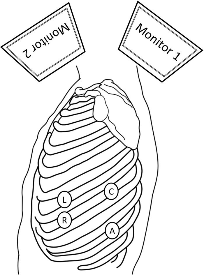
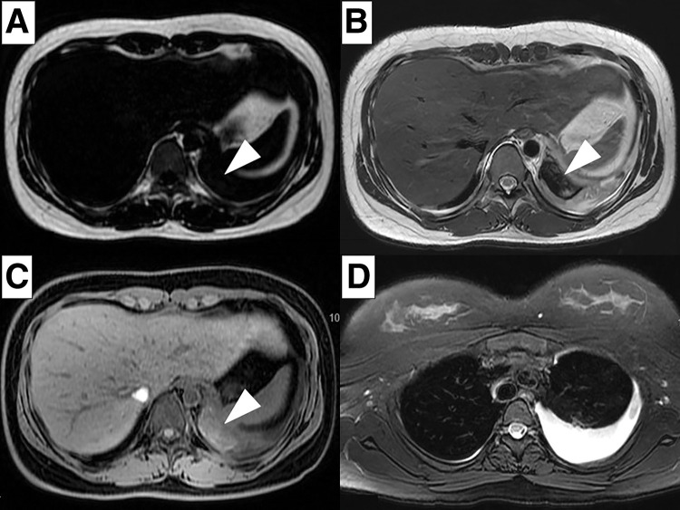
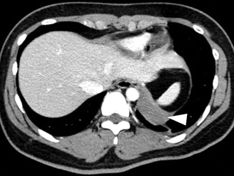
Case presentation: An 18-year-old female presented with a fever and sudden left-sided back pain. Blood tests revealed leukocytosis and an elevated C-reactive protein. Imaging studies identified a well-circumscribed cyst along the left diaphragm, suspected to be an infected bronchogenic cyst. Magnetic resonance imaging 2 days later indicated disease progression with concomitant empyema, prompting emergency surgery. Using the confronting upside-down monitor setting, the cyst was resected. Thoracoscopic findings revealed a dark red cyst and bloody pleural effusion. The surgery was uneventful, and the patient was discharged on postoperative day 2. Bacterial cultures of the pleural effusion and cystic content were negative, and histopathological analysis confirmed the diagnosis of a hemorrhagic bronchogenic cyst.
Conclusions: Hemorrhagic bronchogenic cysts should be considered in the differential diagnosis of intrathoracic cysts presenting with sudden pain. Upfront surgery is recommended for symptomatic bronchogenic cysts, irrespective of the location or etiology. VATS via the confronting upside-down monitor setting is the feasible option alongside the conventional approach.

 求助内容:
求助内容: 应助结果提醒方式:


