Retinal Nerve Fiber Layer Damages from Macular Grasping during Vitrectomy Comparing Traditional and Three-Dimensional Microscope: A Randomized Clinical Trial.
Luigi Caretti, Alberto Guerriero, Giacomo Verzola, Giulia Pillon, Cristina Monterosso, Anna Rita Daniele
{"title":"Retinal Nerve Fiber Layer Damages from Macular Grasping during Vitrectomy Comparing Traditional and Three-Dimensional Microscope: A Randomized Clinical Trial.","authors":"Luigi Caretti, Alberto Guerriero, Giacomo Verzola, Giulia Pillon, Cristina Monterosso, Anna Rita Daniele","doi":"10.4103/joco.joco_214_23","DOIUrl":null,"url":null,"abstract":"<p><strong>Purpose: </strong>To analyze the advantages of the new advanced three-dimensional (3D) heads-up microscopy system in vitreoretinal surgery compared to a standard optical microscope, by evaluating postoperative tissue trauma, defined by the extent of swelling of the arcuate nerve fiber layer (SANFL).</p><p><strong>Methods: </strong>Sixty-four consecutive eyes affected by macular pucker or macular hole underwent macular peeling using a traditional optical microscope (Leica F40) or a 3D heads-up microscopy system (NGENUITY 3D Visualization System). Infrared, blue autofluorescence imaging, and spectral-domain optical coherence tomography (OCT) were performed preoperatively and 2 weeks, 1 month, and 3 months after surgery. The presence of SANFLs was checked postoperatively on infrared and blue autofluorescence fundus imaging, and the extent of each SANFL was measured on OCT near-infrared fundus images.</p><p><strong>Results: </strong>The presence and the extension of SANFLs were comparable in the two groups (<i>P</i> > 0.05), with a regression over time that was similar in the two groups. The change of the peripapillary retinal nerve fiber layer thickness in the various sectors also appeared to have a similar trend with no statistically significant differences in the two groups (<i>P</i> > 0.05).</p><p><strong>Conclusion: </strong>NGENUITY system is comparable in terms of visual and anatomical results, offering perspectives for the integration of new advanced visualization technologies.</p>","PeriodicalId":15423,"journal":{"name":"Journal of Current Ophthalmology","volume":"36 2","pages":"168-175"},"PeriodicalIF":0.9000,"publicationDate":"2025-01-18","publicationTypes":"Journal Article","fieldsOfStudy":null,"isOpenAccess":false,"openAccessPdf":"https://www.ncbi.nlm.nih.gov/pmc/articles/PMC11856124/pdf/","citationCount":"0","resultStr":null,"platform":"Semanticscholar","paperid":null,"PeriodicalName":"Journal of Current Ophthalmology","FirstCategoryId":"1085","ListUrlMain":"https://doi.org/10.4103/joco.joco_214_23","RegionNum":0,"RegionCategory":null,"ArticlePicture":[],"TitleCN":null,"AbstractTextCN":null,"PMCID":null,"EPubDate":"2024/4/1 0:00:00","PubModel":"eCollection","JCR":"Q3","JCRName":"OPHTHALMOLOGY","Score":null,"Total":0}
引用次数: 0
Abstract
Purpose: To analyze the advantages of the new advanced three-dimensional (3D) heads-up microscopy system in vitreoretinal surgery compared to a standard optical microscope, by evaluating postoperative tissue trauma, defined by the extent of swelling of the arcuate nerve fiber layer (SANFL).
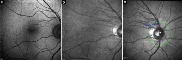
Methods: Sixty-four consecutive eyes affected by macular pucker or macular hole underwent macular peeling using a traditional optical microscope (Leica F40) or a 3D heads-up microscopy system (NGENUITY 3D Visualization System). Infrared, blue autofluorescence imaging, and spectral-domain optical coherence tomography (OCT) were performed preoperatively and 2 weeks, 1 month, and 3 months after surgery. The presence of SANFLs was checked postoperatively on infrared and blue autofluorescence fundus imaging, and the extent of each SANFL was measured on OCT near-infrared fundus images.
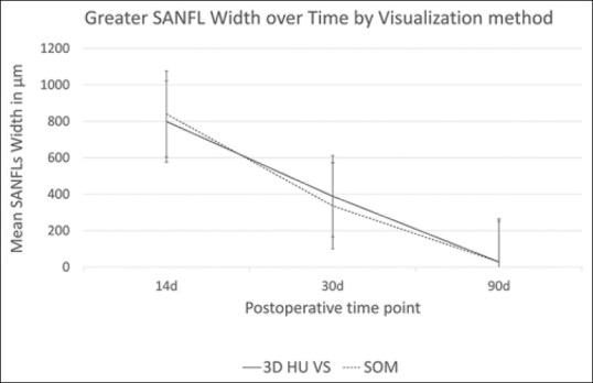
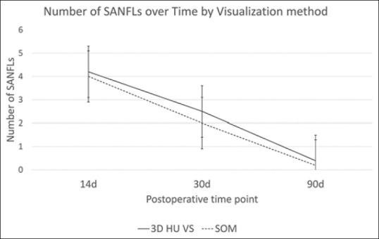
Results: The presence and the extension of SANFLs were comparable in the two groups (P > 0.05), with a regression over time that was similar in the two groups. The change of the peripapillary retinal nerve fiber layer thickness in the various sectors also appeared to have a similar trend with no statistically significant differences in the two groups (P > 0.05).
Conclusion: NGENUITY system is comparable in terms of visual and anatomical results, offering perspectives for the integration of new advanced visualization technologies.
期刊介绍:
Peer Review under the responsibility of Iranian Society of Ophthalmology Journal of Current Ophthalmology, the official publication of the Iranian Society of Ophthalmology, is a peer-reviewed, open-access, scientific journal that welcomes high quality original articles related to vision science and all fields of ophthalmology. Journal of Current Ophthalmology is the continuum of Iranian Journal of Ophthalmology published since 1969.

 求助内容:
求助内容: 应助结果提醒方式:


