Sai Prashanti Chitturi, Vishma Prabhu, Jay Chhablani, Ramesh Venkatesh
{"title":"Progressive Changes in a Torpedo Maculopathy Lesion Over a 6-Year Period.","authors":"Sai Prashanti Chitturi, Vishma Prabhu, Jay Chhablani, Ramesh Venkatesh","doi":"10.4103/joco.joco_216_23","DOIUrl":null,"url":null,"abstract":"<p><strong>Purpose: </strong>To report the longitudinal changes on optical coherence tomography (OCT) in a patient with torpedo maculopathy (TM).</p><p><strong>Methods: </strong>Retrospective observational study.</p><p><strong>Results: </strong>A 37-year-old male, without any ocular symptoms, on dilated fundus examination showed a characteristic torpedo-shaped, flat, horizontally oriented, ovoid-shaped, hypopigmented chorioretinal lesion, temporal to the foveal center. At the most recent visit, after 6 years, the lesion exhibited changes on OCT, including the collapse of the subretinal cleft, the thinning of the retinal pigment epithelium (RPE) layer and inner choroidal layers, and the increased visibility of the underlying choroid and inner retinal excavation. The fundus autofluorescence demonstrated an increase in hypoautofluorescence in the center of the torpedo lesion, which was surrounded by a hyperautofluorescent boundary.</p><p><strong>Conclusion: </strong>This appears to be the first report of longitudinal changes in a classic case of TM, demonstrating changes in the RPE, inner choroidal, and outer retinal layers over time.</p>","PeriodicalId":15423,"journal":{"name":"Journal of Current Ophthalmology","volume":"36 2","pages":"198-200"},"PeriodicalIF":0.9000,"publicationDate":"2025-01-18","publicationTypes":"Journal Article","fieldsOfStudy":null,"isOpenAccess":false,"openAccessPdf":"https://www.ncbi.nlm.nih.gov/pmc/articles/PMC11856115/pdf/","citationCount":"0","resultStr":null,"platform":"Semanticscholar","paperid":null,"PeriodicalName":"Journal of Current Ophthalmology","FirstCategoryId":"1085","ListUrlMain":"https://doi.org/10.4103/joco.joco_216_23","RegionNum":0,"RegionCategory":null,"ArticlePicture":[],"TitleCN":null,"AbstractTextCN":null,"PMCID":null,"EPubDate":"2024/4/1 0:00:00","PubModel":"eCollection","JCR":"Q3","JCRName":"OPHTHALMOLOGY","Score":null,"Total":0}
引用次数: 0
Abstract
Purpose: To report the longitudinal changes on optical coherence tomography (OCT) in a patient with torpedo maculopathy (TM).
Methods: Retrospective observational study.
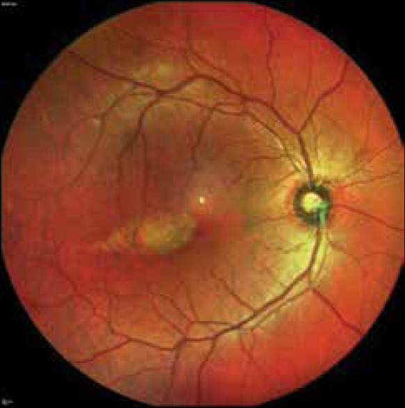
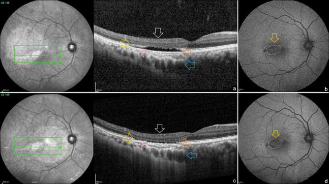
Results: A 37-year-old male, without any ocular symptoms, on dilated fundus examination showed a characteristic torpedo-shaped, flat, horizontally oriented, ovoid-shaped, hypopigmented chorioretinal lesion, temporal to the foveal center. At the most recent visit, after 6 years, the lesion exhibited changes on OCT, including the collapse of the subretinal cleft, the thinning of the retinal pigment epithelium (RPE) layer and inner choroidal layers, and the increased visibility of the underlying choroid and inner retinal excavation. The fundus autofluorescence demonstrated an increase in hypoautofluorescence in the center of the torpedo lesion, which was surrounded by a hyperautofluorescent boundary.
Conclusion: This appears to be the first report of longitudinal changes in a classic case of TM, demonstrating changes in the RPE, inner choroidal, and outer retinal layers over time.
期刊介绍:
Peer Review under the responsibility of Iranian Society of Ophthalmology Journal of Current Ophthalmology, the official publication of the Iranian Society of Ophthalmology, is a peer-reviewed, open-access, scientific journal that welcomes high quality original articles related to vision science and all fields of ophthalmology. Journal of Current Ophthalmology is the continuum of Iranian Journal of Ophthalmology published since 1969.

 求助内容:
求助内容: 应助结果提醒方式:


