Su Ho Bae, Seung Woo Choi, Chang Ki Yoon, Un Chul Park, Kyu Hyung Park, Eun Kyoung Lee
{"title":"Clinical Characteristics and Visual Prognostic Biomarkers in Pericentral Retinitis Pigmentosa: A Study in a South Korean Cohort.","authors":"Su Ho Bae, Seung Woo Choi, Chang Ki Yoon, Un Chul Park, Kyu Hyung Park, Eun Kyoung Lee","doi":"10.3341/kjo.2024.0097","DOIUrl":null,"url":null,"abstract":"<p><strong>Purpose: </strong>To investigate the clinical characteristics of South Korean patients with pericentral retinitis pigmentosa (RP) and to identify clinical biomarkers associated with rapid visual acuity decline based on baseline factors.</p><p><strong>Methods: </strong>This retrospective study included 59 eyes of 31 patients diagnosed with pericentral RP. Comprehensive ophthalmological examinations and genetic sequencing were conducted to assess the baseline characteristics. For biomarker analysis, eyes were categorized into two groups based on the annual rate of change in visual acuity. The clinical findings of the two groups were evaluated to identify the biomarkers associated with rapid loss of visual acuity.</p><p><strong>Results: </strong>Patients with pericentral RP in this study exhibited a mean best-corrected visual acuity of 0.17 ± 0.23 in logarithm of the minimum angle of resolution. The visual field test showed annular or semicircular scotoma with relatively preserved periphery and 27 eyes (45.8%) exhibited no macular complications in optical coherence tomography. Genetic analysis identified genes associated with previous typical and pericentral RP studies but also highlighted that many genetic causes of pericentral RP remain unidentified. Of the 55 eyes for which the rate of visual acuity change could be estimated, 18 exhibited an annual decline of ≥10%, whereas 37 showed an annual decline of <10%. Male sex and prolonged b-wave latency on dark-adapted 0.01 electroretinogram correlated with rapid visual acuity decline in the multivariate analysis.</p><p><strong>Conclusions: </strong>South Korean patients with pericentral RP exhibited a milder phenotype compared to typical RP patients reported in previous studies. Genetic analysis revealed heterogeneity, with mutations in some genes commonly associated with milder forms of RP. Male sex and prolonged b-wave latency on dark-adapted 0.01 electroretinogram were significant biomarkers for predicting rapid visual acuity decline. Monitoring initial b-wave latency is important for predicting visual decline, particularly in male patients with pericentral RP.</p>","PeriodicalId":101356,"journal":{"name":"Korean journal of ophthalmology : KJO","volume":" ","pages":"157-169"},"PeriodicalIF":0.0000,"publicationDate":"2025-04-01","publicationTypes":"Journal Article","fieldsOfStudy":null,"isOpenAccess":false,"openAccessPdf":"https://www.ncbi.nlm.nih.gov/pmc/articles/PMC12010173/pdf/","citationCount":"0","resultStr":null,"platform":"Semanticscholar","paperid":null,"PeriodicalName":"Korean journal of ophthalmology : KJO","FirstCategoryId":"1085","ListUrlMain":"https://doi.org/10.3341/kjo.2024.0097","RegionNum":0,"RegionCategory":null,"ArticlePicture":[],"TitleCN":null,"AbstractTextCN":null,"PMCID":null,"EPubDate":"2025/2/26 0:00:00","PubModel":"Epub","JCR":"","JCRName":"","Score":null,"Total":0}
引用次数: 0
Abstract
Purpose: To investigate the clinical characteristics of South Korean patients with pericentral retinitis pigmentosa (RP) and to identify clinical biomarkers associated with rapid visual acuity decline based on baseline factors.
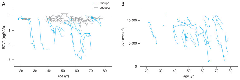
Methods: This retrospective study included 59 eyes of 31 patients diagnosed with pericentral RP. Comprehensive ophthalmological examinations and genetic sequencing were conducted to assess the baseline characteristics. For biomarker analysis, eyes were categorized into two groups based on the annual rate of change in visual acuity. The clinical findings of the two groups were evaluated to identify the biomarkers associated with rapid loss of visual acuity.
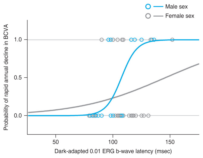
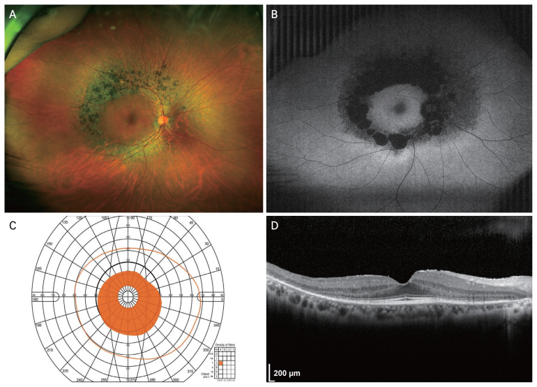
Results: Patients with pericentral RP in this study exhibited a mean best-corrected visual acuity of 0.17 ± 0.23 in logarithm of the minimum angle of resolution. The visual field test showed annular or semicircular scotoma with relatively preserved periphery and 27 eyes (45.8%) exhibited no macular complications in optical coherence tomography. Genetic analysis identified genes associated with previous typical and pericentral RP studies but also highlighted that many genetic causes of pericentral RP remain unidentified. Of the 55 eyes for which the rate of visual acuity change could be estimated, 18 exhibited an annual decline of ≥10%, whereas 37 showed an annual decline of <10%. Male sex and prolonged b-wave latency on dark-adapted 0.01 electroretinogram correlated with rapid visual acuity decline in the multivariate analysis.
Conclusions: South Korean patients with pericentral RP exhibited a milder phenotype compared to typical RP patients reported in previous studies. Genetic analysis revealed heterogeneity, with mutations in some genes commonly associated with milder forms of RP. Male sex and prolonged b-wave latency on dark-adapted 0.01 electroretinogram were significant biomarkers for predicting rapid visual acuity decline. Monitoring initial b-wave latency is important for predicting visual decline, particularly in male patients with pericentral RP.

 求助内容:
求助内容: 应助结果提醒方式:


