Jingjing Zhang, Xiuying Liu, Yinye Huang, Liangyi Kong, Min Su, Zheng Hu
{"title":"3D reconstructed models based on real cervical cancer cases for undergraduate gynecological oncology education: a pre- and post-test study.","authors":"Jingjing Zhang, Xiuying Liu, Yinye Huang, Liangyi Kong, Min Su, Zheng Hu","doi":"10.1186/s41205-025-00256-z","DOIUrl":null,"url":null,"abstract":"<p><strong>Background: </strong>The landscape of medical education is rapidly evolving, driven by advancements in technology. This evolution has ushered in a new era characterized by digitization, connectivity, and intelligence. In this era, traditional teaching methods are being augmented with innovative technologies such as virtual learning, artificial intelligence platforms, and access to cloud-based health platforms. One notable advancement is the integration of three-dimensional (3D) reconstructed models into medical education, particularly in fields like gynecological oncology.</p><p><strong>Methods: </strong>This study introduces 3D reconstructed models based on real cervical cancer cases as a teaching tool for undergraduate gynecological oncology education. Participants were fourth-year Clinical Medicine students of Wuhan University, China. Using student identity document numbers for grouping, half were assigned to the control group (odd numbers) and the other half to the 3D reconstructed model teaching group (even numbers). All the students completed the pre-tests before receiving traditional teaching on cervical intraepithelial lesions and cervical cancer. The control group completed the post-tests after traditional teaching alone, while the 3D reconstructed model teaching group completed the post-tests after receiving the additional 3D reconstructed model teaching. Feedback on this innovative teaching tool was collected. The pre-tests and post-tests covered cervical intraepithelial lesions, cervical cancer and staging system, and female pelvic anatomy.</p><p><strong>Results: </strong>This study includes 267 students, with 134 in the control group and 133 in the 3D reconstructed model teaching group. The pre-test scores of the three tests between the control group and the 3D reconstructed model teaching group showed no statistical difference (p > 0.05). Compared to the control group, the post-test scores of the 3D reconstructed model teaching group in theoretical knowledge of cervical intraepithelial lesions and cervical cancer, female pelvic anatomy and 2018 International Federation of Gynecology and Obstetrics staging system for cervical cancer increased significantly (p < 0.05). Feedback from students underscored the visual benefits and engaging nature of the models, with many expressing that the 3D models provided a clearer representation of cervical cancer and enhanced their learning experience.</p><p><strong>Conclusion: </strong>The integration of 3D reconstructed models into medical education represents a promising approach to address the complexities of teaching intricate subjects in anatomy such as gynecological oncology. These models offer a more intuitive and thorough visualization of anatomical structures and pathological processes, fostering a hands-on and exploratory learning experience for students.</p>","PeriodicalId":72036,"journal":{"name":"3D printing in medicine","volume":"11 1","pages":"7"},"PeriodicalIF":3.1000,"publicationDate":"2025-02-26","publicationTypes":"Journal Article","fieldsOfStudy":null,"isOpenAccess":false,"openAccessPdf":"https://www.ncbi.nlm.nih.gov/pmc/articles/PMC11863587/pdf/","citationCount":"0","resultStr":null,"platform":"Semanticscholar","paperid":null,"PeriodicalName":"3D printing in medicine","FirstCategoryId":"1085","ListUrlMain":"https://doi.org/10.1186/s41205-025-00256-z","RegionNum":0,"RegionCategory":null,"ArticlePicture":[],"TitleCN":null,"AbstractTextCN":null,"PMCID":null,"EPubDate":"","PubModel":"","JCR":"Q1","JCRName":"RADIOLOGY, NUCLEAR MEDICINE & MEDICAL IMAGING","Score":null,"Total":0}
引用次数: 0
Abstract
Background: The landscape of medical education is rapidly evolving, driven by advancements in technology. This evolution has ushered in a new era characterized by digitization, connectivity, and intelligence. In this era, traditional teaching methods are being augmented with innovative technologies such as virtual learning, artificial intelligence platforms, and access to cloud-based health platforms. One notable advancement is the integration of three-dimensional (3D) reconstructed models into medical education, particularly in fields like gynecological oncology.
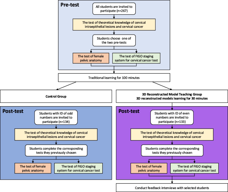
Methods: This study introduces 3D reconstructed models based on real cervical cancer cases as a teaching tool for undergraduate gynecological oncology education. Participants were fourth-year Clinical Medicine students of Wuhan University, China. Using student identity document numbers for grouping, half were assigned to the control group (odd numbers) and the other half to the 3D reconstructed model teaching group (even numbers). All the students completed the pre-tests before receiving traditional teaching on cervical intraepithelial lesions and cervical cancer. The control group completed the post-tests after traditional teaching alone, while the 3D reconstructed model teaching group completed the post-tests after receiving the additional 3D reconstructed model teaching. Feedback on this innovative teaching tool was collected. The pre-tests and post-tests covered cervical intraepithelial lesions, cervical cancer and staging system, and female pelvic anatomy.
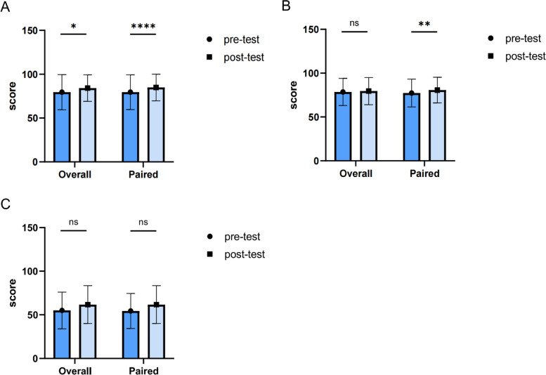
Results: This study includes 267 students, with 134 in the control group and 133 in the 3D reconstructed model teaching group. The pre-test scores of the three tests between the control group and the 3D reconstructed model teaching group showed no statistical difference (p > 0.05). Compared to the control group, the post-test scores of the 3D reconstructed model teaching group in theoretical knowledge of cervical intraepithelial lesions and cervical cancer, female pelvic anatomy and 2018 International Federation of Gynecology and Obstetrics staging system for cervical cancer increased significantly (p < 0.05). Feedback from students underscored the visual benefits and engaging nature of the models, with many expressing that the 3D models provided a clearer representation of cervical cancer and enhanced their learning experience.
Conclusion: The integration of 3D reconstructed models into medical education represents a promising approach to address the complexities of teaching intricate subjects in anatomy such as gynecological oncology. These models offer a more intuitive and thorough visualization of anatomical structures and pathological processes, fostering a hands-on and exploratory learning experience for students.

 求助内容:
求助内容: 应助结果提醒方式:


