Julio Cesar Ramos Cadilho, Paôla Caroline da Silva Mira, Jéssica Peixoto Bem, Penélope Swerts Rodrigues, Francisco José de Queiroz Thomé, Harley Francisco de Oliveira, Fernando Marin Torres, Francisco Wanderley Garcia Paula-Silva, Paulo Nelson-Filho, Alexandra Mussolino de Queiroz
{"title":"Effects of radiation therapy on the morphology and composition of root dentin and cementum in primary teeth.","authors":"Julio Cesar Ramos Cadilho, Paôla Caroline da Silva Mira, Jéssica Peixoto Bem, Penélope Swerts Rodrigues, Francisco José de Queiroz Thomé, Harley Francisco de Oliveira, Fernando Marin Torres, Francisco Wanderley Garcia Paula-Silva, Paulo Nelson-Filho, Alexandra Mussolino de Queiroz","doi":"10.1590/1807-3107bor-2025.vol39.026","DOIUrl":null,"url":null,"abstract":"<p><p>The aim of this study was to evaluate, in vitro, the effects of radiation therapy on the morphology and chemical composition of root dentin and cementum of primary teeth. Roots of human primary teeth were exposed to varying doses of irradiation using a linear accelerator: Group 1 = irradiation dose of up to 30 Gy (n= 6); Group 2 = irradiation dose of up to 42 Gy (n= 6); and Group 3 = irradiation dose of up to 54 Gy (n= 6). Non-irradiated human deciduous teeth were used as controls (n= 3). Energy-dispersive X-ray spectroscopy (EDX) data were analyzed using the chi-square test (alpha = 5%). The morphological evaluation was performed using scanning electron microscopy (SEM). In the cementum, radiation increased inorganic composition and decreased oxygen levels. However, no morphological changes were observed. In the root dentin, obliterated dentinal tubules were observed in specimens irradiated with 54 Gy, with no changes in composition. Thus, radiation therapy significantly altered the morphology and composition of important tooth structures such as dentin and cementum, in primary teeth.</p>","PeriodicalId":9240,"journal":{"name":"Brazilian oral research","volume":"39 ","pages":"e026"},"PeriodicalIF":1.3000,"publicationDate":"2025-02-24","publicationTypes":"Journal Article","fieldsOfStudy":null,"isOpenAccess":false,"openAccessPdf":"https://www.ncbi.nlm.nih.gov/pmc/articles/PMC11849964/pdf/","citationCount":"0","resultStr":null,"platform":"Semanticscholar","paperid":null,"PeriodicalName":"Brazilian oral research","FirstCategoryId":"3","ListUrlMain":"https://doi.org/10.1590/1807-3107bor-2025.vol39.026","RegionNum":4,"RegionCategory":"医学","ArticlePicture":[],"TitleCN":null,"AbstractTextCN":null,"PMCID":null,"EPubDate":"2025/1/1 0:00:00","PubModel":"eCollection","JCR":"Q3","JCRName":"DENTISTRY, ORAL SURGERY & MEDICINE","Score":null,"Total":0}
引用次数: 0
Abstract
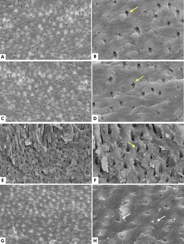
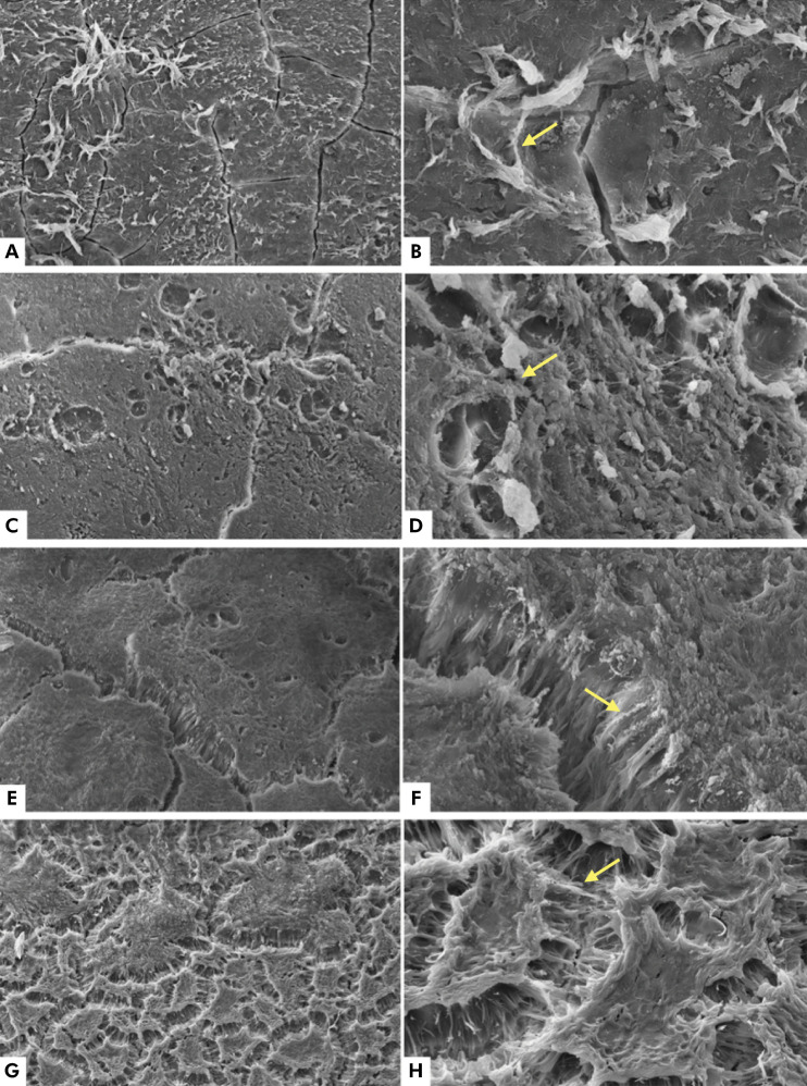
The aim of this study was to evaluate, in vitro, the effects of radiation therapy on the morphology and chemical composition of root dentin and cementum of primary teeth. Roots of human primary teeth were exposed to varying doses of irradiation using a linear accelerator: Group 1 = irradiation dose of up to 30 Gy (n= 6); Group 2 = irradiation dose of up to 42 Gy (n= 6); and Group 3 = irradiation dose of up to 54 Gy (n= 6). Non-irradiated human deciduous teeth were used as controls (n= 3). Energy-dispersive X-ray spectroscopy (EDX) data were analyzed using the chi-square test (alpha = 5%). The morphological evaluation was performed using scanning electron microscopy (SEM). In the cementum, radiation increased inorganic composition and decreased oxygen levels. However, no morphological changes were observed. In the root dentin, obliterated dentinal tubules were observed in specimens irradiated with 54 Gy, with no changes in composition. Thus, radiation therapy significantly altered the morphology and composition of important tooth structures such as dentin and cementum, in primary teeth.
 求助内容:
求助内容: 应助结果提醒方式:


