Israel Muro, Andrea C Qualman, Kenneth Meza Monge, Akshay Pratap, Elizabeth J Kovacs, Juan-Pablo Idrovo
{"title":"Delayed hepatic response and impaired cytokine dynamics in aged mice following burn injury: Implications for elderly patient care.","authors":"Israel Muro, Andrea C Qualman, Kenneth Meza Monge, Akshay Pratap, Elizabeth J Kovacs, Juan-Pablo Idrovo","doi":"10.1371/journal.pone.0316813","DOIUrl":null,"url":null,"abstract":"<p><strong>Introduction: </strong>Burn injuries in elderly patients result in higher morbidity and mortality compared to younger individuals. This study investigates age-related differences in inflammatory hepatic responses to burn injuries.</p><p><strong>Method: </strong>Young (8-10 weeks) and aged (20-21 months) female C57BL/6 mice were subjected to a 15% total body surface area burn or sham injury. Serum and liver samples collected at 3, 6-, 9-, 12-, and 24-hours post-injury were analyzed for serum amyloid A (SAA) levels, SAA1 and SAA2 hepatic gene expression, serum cytokines (IL-6, IL-1β, TNF-α, and IL-10), and hepatic STAT3 activation.</p><p><strong>Results: </strong>Aged mice showed a delayed and dysregulated response. In young mice, SAA levels rose significantly at 6 hours postburn (5.09 ± 0.2-fold), while in aged mice, SAA increased at 12 hours (39.1 ± 2.06-fold), p < 0.01. Hepatic expression of SAA1 and SAA2 also peaked early in young mice (8.357 ± 1.257-fold and 5.91 ± 0.664-fold at 3 hours) but was delayed until 12 hours in aged mice. Young mice demonstrated early IL-6 peaks at 3 hours (990 ± 83.2 pg/ml), while aged mice reached a delayed, higher IL-6 peak at 24 hours (3804 ± 1408 pg/ml, p < 0.05). Similar age-related delays occurred for IL-1β and TNF-α. Aged mice had significantly elevated IL-10 at 6 hours (993.9 ± 99.41 pg/ml vs. 67.69 ± 6.635 pg/ml in young, p < 0.001). STAT3 activation peaked at 3 hours in young mice (2.686 ± 0.226-fold) but was delayed until 24 hours in aged mice (0.5958 ± 0.0368-fold, p < 0.05).</p><p><strong>Conclusions: </strong>This study identifies age-related variations in inflammatory markers and acute hepatic responses to burn injuries, with aged mice showing delayed and reduced inflammatory responses compared to younger counterparts. These findings underscore the importance of age-specific strategies in burn injury management to enhance outcomes for elderly burn patients.</p>","PeriodicalId":20189,"journal":{"name":"PLoS ONE","volume":"20 2","pages":"e0316813"},"PeriodicalIF":2.6000,"publicationDate":"2025-02-24","publicationTypes":"Journal Article","fieldsOfStudy":null,"isOpenAccess":false,"openAccessPdf":"https://www.ncbi.nlm.nih.gov/pmc/articles/PMC11849828/pdf/","citationCount":"0","resultStr":null,"platform":"Semanticscholar","paperid":null,"PeriodicalName":"PLoS ONE","FirstCategoryId":"103","ListUrlMain":"https://doi.org/10.1371/journal.pone.0316813","RegionNum":3,"RegionCategory":"综合性期刊","ArticlePicture":[],"TitleCN":null,"AbstractTextCN":null,"PMCID":null,"EPubDate":"2025/1/1 0:00:00","PubModel":"eCollection","JCR":"Q1","JCRName":"MULTIDISCIPLINARY SCIENCES","Score":null,"Total":0}
引用次数: 0
Abstract
Introduction: Burn injuries in elderly patients result in higher morbidity and mortality compared to younger individuals. This study investigates age-related differences in inflammatory hepatic responses to burn injuries.
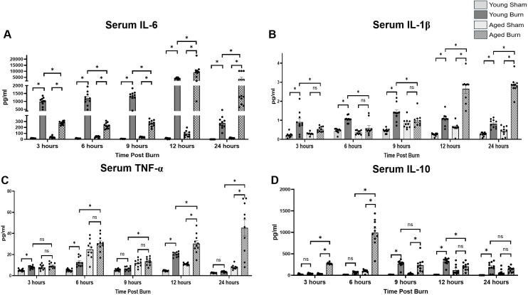
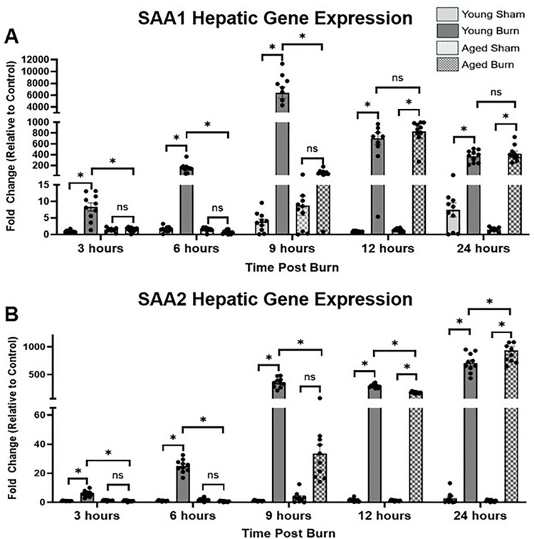
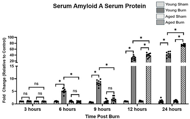
Method: Young (8-10 weeks) and aged (20-21 months) female C57BL/6 mice were subjected to a 15% total body surface area burn or sham injury. Serum and liver samples collected at 3, 6-, 9-, 12-, and 24-hours post-injury were analyzed for serum amyloid A (SAA) levels, SAA1 and SAA2 hepatic gene expression, serum cytokines (IL-6, IL-1β, TNF-α, and IL-10), and hepatic STAT3 activation.
Results: Aged mice showed a delayed and dysregulated response. In young mice, SAA levels rose significantly at 6 hours postburn (5.09 ± 0.2-fold), while in aged mice, SAA increased at 12 hours (39.1 ± 2.06-fold), p < 0.01. Hepatic expression of SAA1 and SAA2 also peaked early in young mice (8.357 ± 1.257-fold and 5.91 ± 0.664-fold at 3 hours) but was delayed until 12 hours in aged mice. Young mice demonstrated early IL-6 peaks at 3 hours (990 ± 83.2 pg/ml), while aged mice reached a delayed, higher IL-6 peak at 24 hours (3804 ± 1408 pg/ml, p < 0.05). Similar age-related delays occurred for IL-1β and TNF-α. Aged mice had significantly elevated IL-10 at 6 hours (993.9 ± 99.41 pg/ml vs. 67.69 ± 6.635 pg/ml in young, p < 0.001). STAT3 activation peaked at 3 hours in young mice (2.686 ± 0.226-fold) but was delayed until 24 hours in aged mice (0.5958 ± 0.0368-fold, p < 0.05).
Conclusions: This study identifies age-related variations in inflammatory markers and acute hepatic responses to burn injuries, with aged mice showing delayed and reduced inflammatory responses compared to younger counterparts. These findings underscore the importance of age-specific strategies in burn injury management to enhance outcomes for elderly burn patients.
期刊介绍:
PLOS ONE is an international, peer-reviewed, open-access, online publication. PLOS ONE welcomes reports on primary research from any scientific discipline. It provides:
* Open-access—freely accessible online, authors retain copyright
* Fast publication times
* Peer review by expert, practicing researchers
* Post-publication tools to indicate quality and impact
* Community-based dialogue on articles
* Worldwide media coverage

 求助内容:
求助内容: 应助结果提醒方式:


