Mohammed Mumuni, Kevin Kofi Adutwum-Ofosu, Benjamin Arko-Boham, Bismarck Afedo Hottor, Nii Koney-Kwaku Koney, Kwame Adu-Bonsaffoh, Samuel Antwi Oppong, Peter Ofori Appiah, John Ahenkorah
{"title":"Histomorphology of placentae of women with sickle cell disease during pregnancy - A case control study.","authors":"Mohammed Mumuni, Kevin Kofi Adutwum-Ofosu, Benjamin Arko-Boham, Bismarck Afedo Hottor, Nii Koney-Kwaku Koney, Kwame Adu-Bonsaffoh, Samuel Antwi Oppong, Peter Ofori Appiah, John Ahenkorah","doi":"10.1371/journal.pone.0319011","DOIUrl":null,"url":null,"abstract":"<p><strong>Background: </strong>Sickle cell disease (SCD) is known to exert multifaceted effects on pregnancy, potentially influencing placental structure and function.</p><p><strong>Aim: </strong>Our aim was to utilize stereology as a precise analytical tool to evaluate the histo-morphologic and functional changes in term placentae of women with SCD against those of non-SCD women.</p><p><strong>Method: </strong>A case control study was conducted at the Korle-Bu Teaching Hospital's labour unit and included 38 pregnant women, comprising 19 cases and 19 controls. Placenta samples were paired and matched with gestational age and taken at term (38 weeks + 2 weeks). Tissue sections were prepared, stained with hematoxylin and eosin, and volume densities of syncytial knots, foetal capillaries, syncytial denuded areas, and intervillous spaces estimated by stereological methods. Statistical analysis was performed to compare mean values between the SCD and control groups.</p><p><strong>Results: </strong>Among the study participants with SCD, 13.16% (5) had sickle cell haemoglobin S (HbSS), 34.21% (13) had haemoglobin C (HbSC) and 2.63% (1) had β-thalassemia (HbS). On stereological assessment, there were statistically significant differences in mean volume densities of syncytial knots (p = < 0.0034), foetal capillaries (p = < 0.0001), syncytial denudations (p = < 0.0028), and intervillous space (p = < 0.0113) between term placentae of women with SCD and those without SCD.</p><p><strong>Conclusion: </strong>SCD placentae may result in a substantial increase in syncytial knot formation, possibly because of hypermaturation of the chorionic villi, significant increase in foetal capillaries potentially due to the hypoxic nature of the SCD placentae, syncytial denuded areas as a result of alteration of the placental syncytium and reduced intervillous spaces which may be due to villous congestion. These findings suggest the need for heightened monitoring of placental function and fetal well-being in pregnancies complicated by SCD to reduce adverse perinatal outcomes.</p>","PeriodicalId":20189,"journal":{"name":"PLoS ONE","volume":"20 2","pages":"e0319011"},"PeriodicalIF":2.6000,"publicationDate":"2025-02-24","publicationTypes":"Journal Article","fieldsOfStudy":null,"isOpenAccess":false,"openAccessPdf":"https://www.ncbi.nlm.nih.gov/pmc/articles/PMC11849815/pdf/","citationCount":"0","resultStr":null,"platform":"Semanticscholar","paperid":null,"PeriodicalName":"PLoS ONE","FirstCategoryId":"103","ListUrlMain":"https://doi.org/10.1371/journal.pone.0319011","RegionNum":3,"RegionCategory":"综合性期刊","ArticlePicture":[],"TitleCN":null,"AbstractTextCN":null,"PMCID":null,"EPubDate":"2025/1/1 0:00:00","PubModel":"eCollection","JCR":"Q1","JCRName":"MULTIDISCIPLINARY SCIENCES","Score":null,"Total":0}
引用次数: 0
Abstract
Background: Sickle cell disease (SCD) is known to exert multifaceted effects on pregnancy, potentially influencing placental structure and function.
Aim: Our aim was to utilize stereology as a precise analytical tool to evaluate the histo-morphologic and functional changes in term placentae of women with SCD against those of non-SCD women.
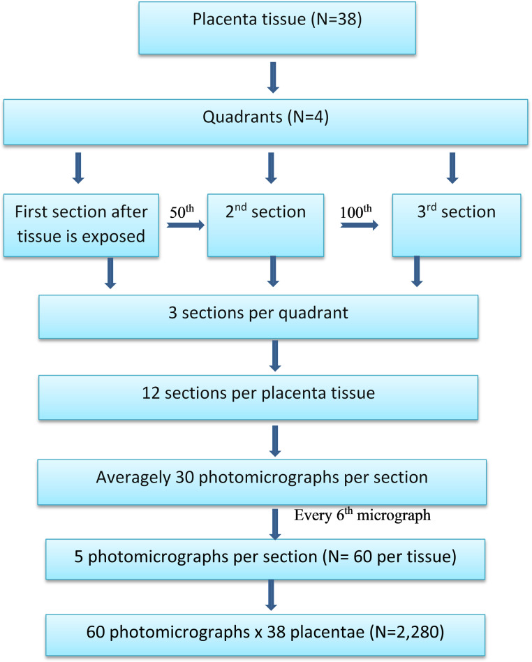
Method: A case control study was conducted at the Korle-Bu Teaching Hospital's labour unit and included 38 pregnant women, comprising 19 cases and 19 controls. Placenta samples were paired and matched with gestational age and taken at term (38 weeks + 2 weeks). Tissue sections were prepared, stained with hematoxylin and eosin, and volume densities of syncytial knots, foetal capillaries, syncytial denuded areas, and intervillous spaces estimated by stereological methods. Statistical analysis was performed to compare mean values between the SCD and control groups.
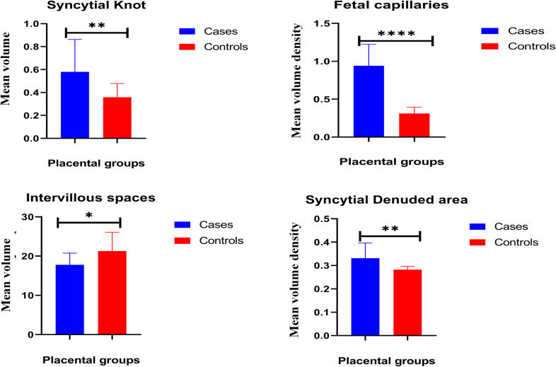
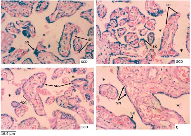
Results: Among the study participants with SCD, 13.16% (5) had sickle cell haemoglobin S (HbSS), 34.21% (13) had haemoglobin C (HbSC) and 2.63% (1) had β-thalassemia (HbS). On stereological assessment, there were statistically significant differences in mean volume densities of syncytial knots (p = < 0.0034), foetal capillaries (p = < 0.0001), syncytial denudations (p = < 0.0028), and intervillous space (p = < 0.0113) between term placentae of women with SCD and those without SCD.
Conclusion: SCD placentae may result in a substantial increase in syncytial knot formation, possibly because of hypermaturation of the chorionic villi, significant increase in foetal capillaries potentially due to the hypoxic nature of the SCD placentae, syncytial denuded areas as a result of alteration of the placental syncytium and reduced intervillous spaces which may be due to villous congestion. These findings suggest the need for heightened monitoring of placental function and fetal well-being in pregnancies complicated by SCD to reduce adverse perinatal outcomes.
期刊介绍:
PLOS ONE is an international, peer-reviewed, open-access, online publication. PLOS ONE welcomes reports on primary research from any scientific discipline. It provides:
* Open-access—freely accessible online, authors retain copyright
* Fast publication times
* Peer review by expert, practicing researchers
* Post-publication tools to indicate quality and impact
* Community-based dialogue on articles
* Worldwide media coverage

 求助内容:
求助内容: 应助结果提醒方式:


