Joe Chin-Hun Kuo, Marshall J. Colville, Michelle R. Sorkin, Jacky Lok Ka Kuo, Ling Ting Huang, Dana N. Thornlow, Gwendolyn M. Beacham, Gunther Hollopeter, Matthew P. DeLisa, Christopher A. Alabi* and Matthew J. Paszek*,
{"title":"Bio-orthogonal Glycan Imaging of Cultured Cells and Whole Animal C. elegans with Expansion Microscopy","authors":"Joe Chin-Hun Kuo, Marshall J. Colville, Michelle R. Sorkin, Jacky Lok Ka Kuo, Ling Ting Huang, Dana N. Thornlow, Gwendolyn M. Beacham, Gunther Hollopeter, Matthew P. DeLisa, Christopher A. Alabi* and Matthew J. Paszek*, ","doi":"10.1021/acscentsci.4c0106110.1021/acscentsci.4c01061","DOIUrl":null,"url":null,"abstract":"<p >Complex carbohydrates called glycans play crucial roles in regulating cell and tissue physiology, but how they map to nanoscale anatomical features must still be resolved. Here, we present the first nanoscale map of mucin-type <i>O</i>-glycans throughout the entirety of the <i>Caenorhabditis elegans</i> model organism. We constructed a library of multifunctional linkers to probe and anchor metabolically labeled glycans in expansion microscopy (ExM). A flexible strategy was demonstrated for the chemical synthesis of linkers with a broad inventory of bio-orthogonal functional groups, fluorophores, anchorage chemistries, and linker arms. Employing <i>C. elegans</i> as a test bed, metabolically labeled <i>O</i>-glycans were resolved on the gut microvilli and other nanoscale anatomical features. Transmission electron microscopy images of <i>C. elegans</i> nanoanatomy validated the fidelity and isotropy of gel expansion. Whole organism maps of <i>C. elegans O</i>-glycosylation in the first larval stage revealed <i>O</i>-glycan “hotspots” in unexpected anatomical locations, including the body wall furrows. Beyond <i>C. elegans</i>, we validated ExM protocols for nanoscale imaging of metabolically labeled glycans on cultured mammalian cells. Together, our results suggest the broad applicability of the multifunctional reagents for imaging glycans and other metabolically labeled biomolecules at enhanced resolutions with ExM.</p><p >Specialized chemical linkers embed molecules mimicking natural metabolites into an expandable gel, including cell surface sugars known as glycans, and light up nanostructures across entire nematodes.</p>","PeriodicalId":10,"journal":{"name":"ACS Central Science","volume":"11 2","pages":"193–207 193–207"},"PeriodicalIF":12.7000,"publicationDate":"2024-11-23","publicationTypes":"Journal Article","fieldsOfStudy":null,"isOpenAccess":false,"openAccessPdf":"https://pubs.acs.org/doi/epdf/10.1021/acscentsci.4c01061","citationCount":"0","resultStr":null,"platform":"Semanticscholar","paperid":null,"PeriodicalName":"ACS Central Science","FirstCategoryId":"92","ListUrlMain":"https://pubs.acs.org/doi/10.1021/acscentsci.4c01061","RegionNum":1,"RegionCategory":"化学","ArticlePicture":[],"TitleCN":null,"AbstractTextCN":null,"PMCID":null,"EPubDate":"","PubModel":"","JCR":"Q1","JCRName":"CHEMISTRY, MULTIDISCIPLINARY","Score":null,"Total":0}
引用次数: 0
Abstract
Complex carbohydrates called glycans play crucial roles in regulating cell and tissue physiology, but how they map to nanoscale anatomical features must still be resolved. Here, we present the first nanoscale map of mucin-type O-glycans throughout the entirety of the Caenorhabditis elegans model organism. We constructed a library of multifunctional linkers to probe and anchor metabolically labeled glycans in expansion microscopy (ExM). A flexible strategy was demonstrated for the chemical synthesis of linkers with a broad inventory of bio-orthogonal functional groups, fluorophores, anchorage chemistries, and linker arms. Employing C. elegans as a test bed, metabolically labeled O-glycans were resolved on the gut microvilli and other nanoscale anatomical features. Transmission electron microscopy images of C. elegans nanoanatomy validated the fidelity and isotropy of gel expansion. Whole organism maps of C. elegans O-glycosylation in the first larval stage revealed O-glycan “hotspots” in unexpected anatomical locations, including the body wall furrows. Beyond C. elegans, we validated ExM protocols for nanoscale imaging of metabolically labeled glycans on cultured mammalian cells. Together, our results suggest the broad applicability of the multifunctional reagents for imaging glycans and other metabolically labeled biomolecules at enhanced resolutions with ExM.
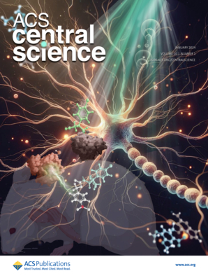
Specialized chemical linkers embed molecules mimicking natural metabolites into an expandable gel, including cell surface sugars known as glycans, and light up nanostructures across entire nematodes.
期刊介绍:
ACS Central Science publishes significant primary reports on research in chemistry and allied fields where chemical approaches are pivotal. As the first fully open-access journal by the American Chemical Society, it covers compelling and important contributions to the broad chemistry and scientific community. "Central science," a term popularized nearly 40 years ago, emphasizes chemistry's central role in connecting physical and life sciences, and fundamental sciences with applied disciplines like medicine and engineering. The journal focuses on exceptional quality articles, addressing advances in fundamental chemistry and interdisciplinary research.
 求助内容:
求助内容: 应助结果提醒方式:


