Eleftheria Michalaki, Alexis N Pulliam, Pooja M Datta Roy, J Brandon Dixon, Michelle C LaPlaca
{"title":"Near-Infrared Imaging of Glymphatic Clearance in a Pre-Clinical Model of Repetitive Closed Head Traumatic Brain Injury.","authors":"Eleftheria Michalaki, Alexis N Pulliam, Pooja M Datta Roy, J Brandon Dixon, Michelle C LaPlaca","doi":"10.1089/neur.2024.0128","DOIUrl":null,"url":null,"abstract":"<p><p>Traumatic brain injury (TBI) is a major health disorder for which there are few treatments. The glymphatic system is the brain's inbuilt lymphatic-like system that is thought to be responsible for clearing waste products from the brain to the lymph nodes. Although there is evidence that glymphatic drainage is crucial for brain homeostasis, its role in TBI pathogenesis remains elusive. Here, we investigated how glymphatic clearance is altered following TBI in rats using real-time non-invasive imaging. Twenty-four hours following repetitive closed-head TBI or sham conditions, we injected infrared dye intraventricularly and used near-infrared (NIR) imaging to quantify signal intensity, intensity over time, and appearance time of NIR dye in different brain regions. TBI yielded a lower NIR signal and lower rate of NIR dye change in the lateral ventricle and surrounding parietal cortex compared with sham conditions, indicating reduced cerebrospinal fluid perfusion. NIR dye appearance took significantly longer to reach the anterior regions of the brain, while perfusion to the posterior of the brain was faster in TBI compared with sham animals. Aquaporin-4 (AQP4) expression was reduced 24 h after TBI across all cortical regions examined in the posterior of the brain and in the ventral cortex at all coronal levels, suggesting a complex relationship between AQP4 and glymph function. Furthermore, NIR imaging revealed that NIR dye was detectable in the cervical lymph nodes (CLNs) of sham animals but not in TBI animals, yet there was evidence of blood accumulation in the CLNs of TBI animals, suggesting that TBI-related extravascular blood is removed through the glymph system. These data indicate that TBI disrupts normal brain efflux kinetics and reduces glymphatic drainage to the CLNs, demonstrating that restoring glymphatic function may be a promising therapeutic target.</p>","PeriodicalId":74300,"journal":{"name":"Neurotrauma reports","volume":"6 1","pages":"115-128"},"PeriodicalIF":1.8000,"publicationDate":"2025-01-30","publicationTypes":"Journal Article","fieldsOfStudy":null,"isOpenAccess":false,"openAccessPdf":"https://www.ncbi.nlm.nih.gov/pmc/articles/PMC11839536/pdf/","citationCount":"0","resultStr":null,"platform":"Semanticscholar","paperid":null,"PeriodicalName":"Neurotrauma reports","FirstCategoryId":"1085","ListUrlMain":"https://doi.org/10.1089/neur.2024.0128","RegionNum":0,"RegionCategory":null,"ArticlePicture":[],"TitleCN":null,"AbstractTextCN":null,"PMCID":null,"EPubDate":"2025/1/1 0:00:00","PubModel":"eCollection","JCR":"Q3","JCRName":"CLINICAL NEUROLOGY","Score":null,"Total":0}
引用次数: 0
Abstract
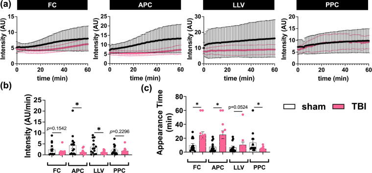
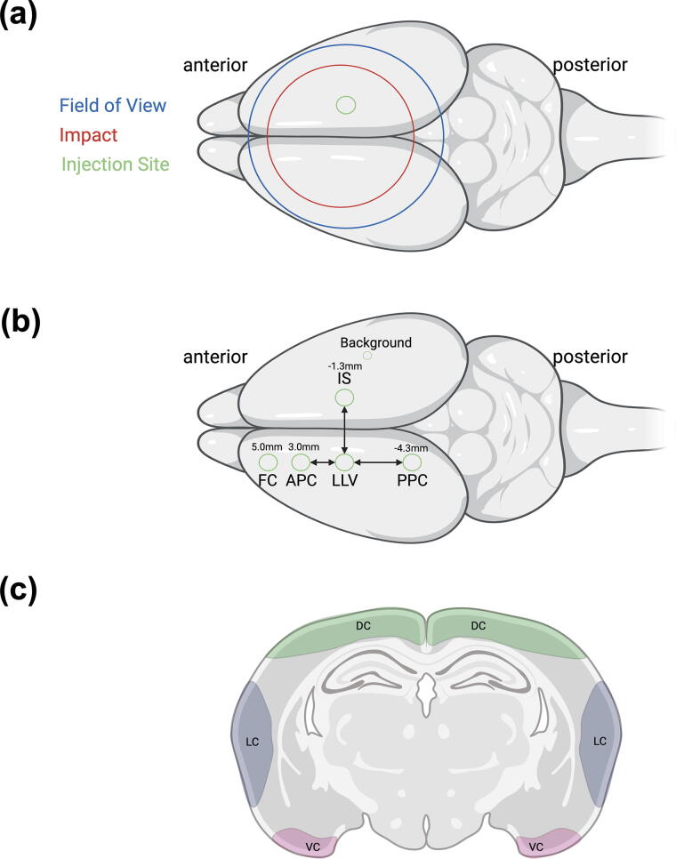
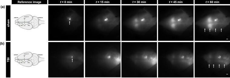
Traumatic brain injury (TBI) is a major health disorder for which there are few treatments. The glymphatic system is the brain's inbuilt lymphatic-like system that is thought to be responsible for clearing waste products from the brain to the lymph nodes. Although there is evidence that glymphatic drainage is crucial for brain homeostasis, its role in TBI pathogenesis remains elusive. Here, we investigated how glymphatic clearance is altered following TBI in rats using real-time non-invasive imaging. Twenty-four hours following repetitive closed-head TBI or sham conditions, we injected infrared dye intraventricularly and used near-infrared (NIR) imaging to quantify signal intensity, intensity over time, and appearance time of NIR dye in different brain regions. TBI yielded a lower NIR signal and lower rate of NIR dye change in the lateral ventricle and surrounding parietal cortex compared with sham conditions, indicating reduced cerebrospinal fluid perfusion. NIR dye appearance took significantly longer to reach the anterior regions of the brain, while perfusion to the posterior of the brain was faster in TBI compared with sham animals. Aquaporin-4 (AQP4) expression was reduced 24 h after TBI across all cortical regions examined in the posterior of the brain and in the ventral cortex at all coronal levels, suggesting a complex relationship between AQP4 and glymph function. Furthermore, NIR imaging revealed that NIR dye was detectable in the cervical lymph nodes (CLNs) of sham animals but not in TBI animals, yet there was evidence of blood accumulation in the CLNs of TBI animals, suggesting that TBI-related extravascular blood is removed through the glymph system. These data indicate that TBI disrupts normal brain efflux kinetics and reduces glymphatic drainage to the CLNs, demonstrating that restoring glymphatic function may be a promising therapeutic target.

 求助内容:
求助内容: 应助结果提醒方式:


