{"title":"Dynamic feature of infrapatellar fat pad during walking in patients with knee osteoarthritis.","authors":"Miharu Sugimoto, Yosuke Ishii, Yuko Nakashima, Goki Kamei, Akinori Nekomoto, Takato Hashizume, Riko Okinaka, Kohei Matsumura, Makoto Takahashi, Nobuo Adachi","doi":"10.1007/s10396-025-01518-3","DOIUrl":null,"url":null,"abstract":"<p><strong>Purpose: </strong>The infrapatellar fat pad (IFP) absorbs mechanical stress in the knee joint owing to flexible morphological changes. The IFP is a key factor in knee osteoarthritis (OA); however, its dynamic feature during walking remains unknown. This study aimed to investigate whether the morphological changes in the IFP during walking involve specific features for patients with knee OA.</p><p><strong>Methods: </strong>Twelve patients with knee OA, 12 healthy young volunteers, and 12 healthy elderly volunteers were recruited. The IFP was evaluated using ultrasonography, and dynamics were recorded in video mode during walking. The IFP value was identified as the thickness between the patellar tendon and proximal tibial line. The morphological change in the IFP (ΔIFP) was shown as the difference in IFP value between maximum and at initial contact on the waveform. Kinematics and kinetics data were evaluated using a three-dimensional motion analysis system synchronized with ultrasonography, and the knee flexion angle and its moment in the stance phase were evaluated.</p><p><strong>Results: </strong>In the patients with knee OA, the ΔIFP was lower than that in healthy volunteers, but there was no difference between control groups (knee OA: 1.4 ± 0.3 mm, elderly control: 1.8 ± 0.2 mm, young control: 2.1 ± 0.5 mm, p < 0.05). In all the groups, there was no significant correlation between the IFP values and kinetic parameters, including the range of knee flexion angles and gait speed.</p><p><strong>Conclusion: </strong>Insufficient morphological changes in the IFP during walking could be a feature of knee OA.</p>","PeriodicalId":50130,"journal":{"name":"Journal of Medical Ultrasonics","volume":" ","pages":"219-226"},"PeriodicalIF":2.1000,"publicationDate":"2025-04-01","publicationTypes":"Journal Article","fieldsOfStudy":null,"isOpenAccess":false,"openAccessPdf":"https://www.ncbi.nlm.nih.gov/pmc/articles/PMC12018517/pdf/","citationCount":"0","resultStr":null,"platform":"Semanticscholar","paperid":null,"PeriodicalName":"Journal of Medical Ultrasonics","FirstCategoryId":"3","ListUrlMain":"https://doi.org/10.1007/s10396-025-01518-3","RegionNum":4,"RegionCategory":"医学","ArticlePicture":[],"TitleCN":null,"AbstractTextCN":null,"PMCID":null,"EPubDate":"2025/2/22 0:00:00","PubModel":"Epub","JCR":"Q3","JCRName":"RADIOLOGY, NUCLEAR MEDICINE & MEDICAL IMAGING","Score":null,"Total":0}
引用次数: 0
Abstract
Purpose: The infrapatellar fat pad (IFP) absorbs mechanical stress in the knee joint owing to flexible morphological changes. The IFP is a key factor in knee osteoarthritis (OA); however, its dynamic feature during walking remains unknown. This study aimed to investigate whether the morphological changes in the IFP during walking involve specific features for patients with knee OA.
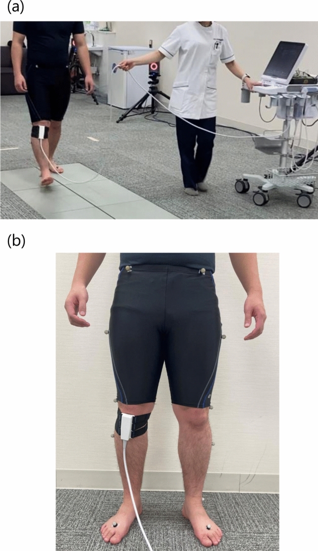
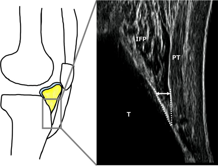
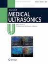
Methods: Twelve patients with knee OA, 12 healthy young volunteers, and 12 healthy elderly volunteers were recruited. The IFP was evaluated using ultrasonography, and dynamics were recorded in video mode during walking. The IFP value was identified as the thickness between the patellar tendon and proximal tibial line. The morphological change in the IFP (ΔIFP) was shown as the difference in IFP value between maximum and at initial contact on the waveform. Kinematics and kinetics data were evaluated using a three-dimensional motion analysis system synchronized with ultrasonography, and the knee flexion angle and its moment in the stance phase were evaluated.
Results: In the patients with knee OA, the ΔIFP was lower than that in healthy volunteers, but there was no difference between control groups (knee OA: 1.4 ± 0.3 mm, elderly control: 1.8 ± 0.2 mm, young control: 2.1 ± 0.5 mm, p < 0.05). In all the groups, there was no significant correlation between the IFP values and kinetic parameters, including the range of knee flexion angles and gait speed.
Conclusion: Insufficient morphological changes in the IFP during walking could be a feature of knee OA.
期刊介绍:
The Journal of Medical Ultrasonics is the official journal of the Japan Society of Ultrasonics in Medicine. The main purpose of the journal is to provide forum for the publication of papers documenting recent advances and new developments in the entire field of ultrasound in medicine and biology, encompassing both the medical and the engineering aspects of the science.The journal welcomes original articles, review articles, images, and letters to the editor.The journal also provides state-of-the-art information such as announcements from the boards and the committees of the society.

 求助内容:
求助内容: 应助结果提醒方式:


