Germán Alfredo Gutiérrez-Liberato, Mélanie Duc, Vytautas Eigirdas, Carolina Romeiro Fernandes Chagas
{"title":"Leucocytozoon infections in tits (Aves, Paridae): blood and tissue stages investigated using an integrative approach.","authors":"Germán Alfredo Gutiérrez-Liberato, Mélanie Duc, Vytautas Eigirdas, Carolina Romeiro Fernandes Chagas","doi":"10.1051/parasite/2025007","DOIUrl":null,"url":null,"abstract":"<p><p>Leucocytozoon species are cosmopolitan and prevalent avian parasites, with some infections being lethal, mainly due to the exo-erythrocytic development of the parasite in bird tissues. The patterns of exo-erythrocytic development in Leucocytozoon spp. infections in wild birds remain poorly studied. This study investigated the development of Leucocytozoon spp. tissue stages in tits (Paridae). Great tits (Parus major), Blue tits (Cyanistes caeruleus), and Coal tits (Periparus ater) were screened for infections using an integrative approach that consisted of microscopic analysis of thin blood smears, histological techniques, chromogenic in situ hybridization (CISH), PCR-based methods, and phylogenetic analysis. In total, 41 individuals were analyzed (eight naturally infected that were selected and euthanized, and 33 found dead in the wild and opportunistically sampled). Among the naturally infected birds, all individuals that were microscopically positive for Leucocytozoon species were also PCR-positive for these parasites. Co-infections with Plasmodium spp. and Haemoproteus spp. were commonly found, mainly among the opportunistically sampled birds. Two morphotypes were identified, Leucocytozoon majoris (Laveran, 1902) and Leucocytozoon fringillinarum Woodcock, 1910. Tissue stages were present in three birds sampled exclusively during the non-breeding season, two of them with meronts developing in the kidneys and liver, and one individual with a megalomeront in the heart. All the exo-erythrocytic stages were confirmed to be Leucocytozoon spp. by CISH using a Leucocytozoon genus-specific probe. Phylogenetic analysis placed parasite lineages with different morphotypes in separate clades. The developmental patterns of exo-erythrocytic stages of Leucocytozoon spp. in naturally infected passerines are poorly understood, requiring further research.</p>","PeriodicalId":19796,"journal":{"name":"Parasite","volume":"32 ","pages":"13"},"PeriodicalIF":2.4000,"publicationDate":"2025-01-01","publicationTypes":"Journal Article","fieldsOfStudy":null,"isOpenAccess":false,"openAccessPdf":"https://www.ncbi.nlm.nih.gov/pmc/articles/PMC11843983/pdf/","citationCount":"0","resultStr":null,"platform":"Semanticscholar","paperid":null,"PeriodicalName":"Parasite","FirstCategoryId":"3","ListUrlMain":"https://doi.org/10.1051/parasite/2025007","RegionNum":2,"RegionCategory":"医学","ArticlePicture":[],"TitleCN":null,"AbstractTextCN":null,"PMCID":null,"EPubDate":"2025/2/21 0:00:00","PubModel":"Epub","JCR":"Q2","JCRName":"PARASITOLOGY","Score":null,"Total":0}
引用次数: 0
Abstract
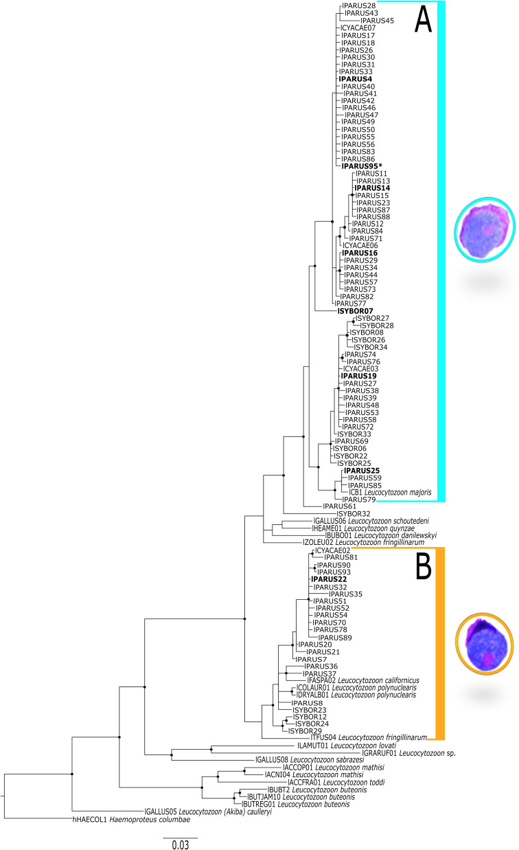
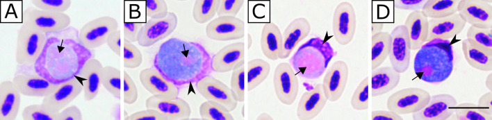
Leucocytozoon species are cosmopolitan and prevalent avian parasites, with some infections being lethal, mainly due to the exo-erythrocytic development of the parasite in bird tissues. The patterns of exo-erythrocytic development in Leucocytozoon spp. infections in wild birds remain poorly studied. This study investigated the development of Leucocytozoon spp. tissue stages in tits (Paridae). Great tits (Parus major), Blue tits (Cyanistes caeruleus), and Coal tits (Periparus ater) were screened for infections using an integrative approach that consisted of microscopic analysis of thin blood smears, histological techniques, chromogenic in situ hybridization (CISH), PCR-based methods, and phylogenetic analysis. In total, 41 individuals were analyzed (eight naturally infected that were selected and euthanized, and 33 found dead in the wild and opportunistically sampled). Among the naturally infected birds, all individuals that were microscopically positive for Leucocytozoon species were also PCR-positive for these parasites. Co-infections with Plasmodium spp. and Haemoproteus spp. were commonly found, mainly among the opportunistically sampled birds. Two morphotypes were identified, Leucocytozoon majoris (Laveran, 1902) and Leucocytozoon fringillinarum Woodcock, 1910. Tissue stages were present in three birds sampled exclusively during the non-breeding season, two of them with meronts developing in the kidneys and liver, and one individual with a megalomeront in the heart. All the exo-erythrocytic stages were confirmed to be Leucocytozoon spp. by CISH using a Leucocytozoon genus-specific probe. Phylogenetic analysis placed parasite lineages with different morphotypes in separate clades. The developmental patterns of exo-erythrocytic stages of Leucocytozoon spp. in naturally infected passerines are poorly understood, requiring further research.
期刊介绍:
Parasite is an international open-access, peer-reviewed, online journal publishing high quality papers on all aspects of human and animal parasitology. Reviews, articles and short notes may be submitted. Fields include, but are not limited to: general, medical and veterinary parasitology; morphology, including ultrastructure; parasite systematics, including entomology, acarology, helminthology and protistology, and molecular analyses; molecular biology and biochemistry; immunology of parasitic diseases; host-parasite relationships; ecology and life history of parasites; epidemiology; therapeutics; new diagnostic tools.
All papers in Parasite are published in English. Manuscripts should have a broad interest and must not have been published or submitted elsewhere. No limit is imposed on the length of manuscripts, but they should be concisely written. Papers of limited interest such as case reports, epidemiological studies in punctual areas, isolated new geographical records, and systematic descriptions of single species will generally not be accepted, but might be considered if the authors succeed in demonstrating their interest.
 求助内容:
求助内容: 应助结果提醒方式:


