{"title":"Development of Macular Atrophy after Macular Hole Surgery in an Eye with Retinitis Pigmentosa.","authors":"Yuki Goto, Kazuki Kuniyoshi, Kensuke Goto, Tomoyasu Kayazawa, Taro Kominami, Fukutaro Mano, Masuo Sakamoto, Chiharu Iwahashi, Shunji Kusaka","doi":"10.1159/000543599","DOIUrl":null,"url":null,"abstract":"<p><strong>Introduction: </strong>Macular hole is a rare complication in patients with retinitis pigmentosa that significantly reduces visual acuity. Although vitreous surgery for macular holes generally yields favorable outcomes, postoperative macular atrophy has been reported. We report the second case of retinitis pigmentosa in a patient who developed a 13-year progressive macular atrophy after macular hole surgery.</p><p><strong>Case presentation: </strong>A 64-year-old Japanese woman, who had been diagnosed with retinitis pigmentosa at 52 years of age, presented to our hospital with blurred vision in her left eye. Phacovitrectomy of the left eye was performed after a full-thickness macular hole was revealed by optical coherence tomography. We stained the internal limiting membrane during surgery using 0.05% indocyanine green and peeled it around the macular hole. Nevertheless, slight atrophy of the retinal pigment epithelium appeared in the left macula 17 days after surgery. The macular hole closed 1 year after surgery, and the macular atrophy gradually became more apparent and enlarged. Thirteen years later, atrophy had expanded to 2.5-disc diameters, and the left decimal best-corrected visual acuity was 0.1; no macular degeneration appeared in the right eye. Genetic examination revealed compound heterozygous variants in the <i>EYS</i> gene.</p><p><strong>Conclusion: </strong>Macular atrophy can develop after dye-assisted macular hole surgery for patients with retinitis pigmentosa. Potential risk factors for the development of postoperative macular atrophy include dye toxicity, light toxicity, surgical intervention in the macula, postoperative inflammation, and genotype. However, the exact cause of atrophy remains uncertain.</p>","PeriodicalId":9635,"journal":{"name":"Case Reports in Ophthalmology","volume":"16 1","pages":"107-113"},"PeriodicalIF":0.6000,"publicationDate":"2025-01-17","publicationTypes":"Journal Article","fieldsOfStudy":null,"isOpenAccess":false,"openAccessPdf":"https://www.ncbi.nlm.nih.gov/pmc/articles/PMC11842103/pdf/","citationCount":"0","resultStr":null,"platform":"Semanticscholar","paperid":null,"PeriodicalName":"Case Reports in Ophthalmology","FirstCategoryId":"1085","ListUrlMain":"https://doi.org/10.1159/000543599","RegionNum":0,"RegionCategory":null,"ArticlePicture":[],"TitleCN":null,"AbstractTextCN":null,"PMCID":null,"EPubDate":"2025/1/1 0:00:00","PubModel":"eCollection","JCR":"Q4","JCRName":"OPHTHALMOLOGY","Score":null,"Total":0}
引用次数: 0
Abstract
Introduction: Macular hole is a rare complication in patients with retinitis pigmentosa that significantly reduces visual acuity. Although vitreous surgery for macular holes generally yields favorable outcomes, postoperative macular atrophy has been reported. We report the second case of retinitis pigmentosa in a patient who developed a 13-year progressive macular atrophy after macular hole surgery.
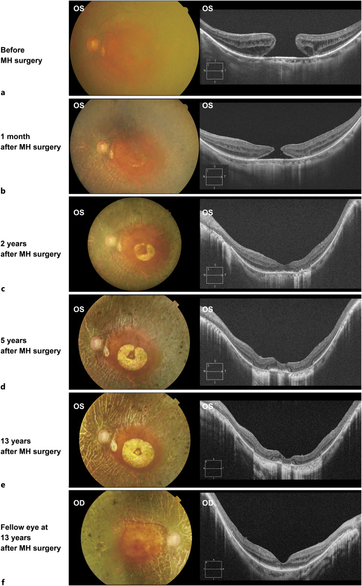
Case presentation: A 64-year-old Japanese woman, who had been diagnosed with retinitis pigmentosa at 52 years of age, presented to our hospital with blurred vision in her left eye. Phacovitrectomy of the left eye was performed after a full-thickness macular hole was revealed by optical coherence tomography. We stained the internal limiting membrane during surgery using 0.05% indocyanine green and peeled it around the macular hole. Nevertheless, slight atrophy of the retinal pigment epithelium appeared in the left macula 17 days after surgery. The macular hole closed 1 year after surgery, and the macular atrophy gradually became more apparent and enlarged. Thirteen years later, atrophy had expanded to 2.5-disc diameters, and the left decimal best-corrected visual acuity was 0.1; no macular degeneration appeared in the right eye. Genetic examination revealed compound heterozygous variants in the EYS gene.
Conclusion: Macular atrophy can develop after dye-assisted macular hole surgery for patients with retinitis pigmentosa. Potential risk factors for the development of postoperative macular atrophy include dye toxicity, light toxicity, surgical intervention in the macula, postoperative inflammation, and genotype. However, the exact cause of atrophy remains uncertain.
期刊介绍:
This peer-reviewed online-only journal publishes original case reports covering the entire spectrum of ophthalmology, including prevention, diagnosis, treatment, toxicities of therapy, supportive care, quality-of-life, and survivorship issues. The submission of negative results is strongly encouraged. The journal will also accept case reports dealing with the use of novel technologies, both in the arena of diagnosis and treatment. Supplementary material is welcomed. The intent of the journal is to provide clinicians and researchers with a tool to disseminate their personal experiences to a wider public as well as to review interesting cases encountered by colleagues all over the world. Universally used terms can be searched across the entire growing collection of case reports, further facilitating the retrieval of specific information. Following the open access principle, the entire contents can be retrieved at no charge, guaranteeing easy access to this valuable source of anecdotal information at all times.

 求助内容:
求助内容: 应助结果提醒方式:


