Sumer Doctor, Ayushi Agarwal, Mohammad Javed Ali, Nandini Bothra
{"title":"Dentigerous Cyst Masquerading as Lacrimal Sac Diverticulitis.","authors":"Sumer Doctor, Ayushi Agarwal, Mohammad Javed Ali, Nandini Bothra","doi":"10.1159/000543279","DOIUrl":null,"url":null,"abstract":"<p><strong>Introduction: </strong>Dentigerous cysts (DCs) involving the orbit are extremely rare. The authors report a unique case of stand-alone orbital DC associated with ectopic canine tooth masquerading as a lacrimal sac diverticulitis with superadded preseptal cellulitis.</p><p><strong>Case presentation: </strong>A 21-year-old lady presented with left inferomedial swelling, associated with pain and redness of 1-week-duration. A lacrimal sac diverticulitis with preseptal cellulitis was suspected and imaging was requested in view of progression. Computed tomography scan of orbit revealed a well-defined heterogenous mass, with associated hyperdense lesion within the mass, prompting a possibility of a foreign body granuloma. Surgical exploration and excision confirmed the diagnosis of an ectopic canine tooth with associated DC along with excessive adhesion of the cyst to the lacrimal sac wall. Maxillary sinus did not show any abnormality. Lacrimal irrigation was patent with no recurrence noted at 6-month follow-up.</p><p><strong>Conclusion: </strong>This report highlights the presentation of DC with associated ectopic canine tooth as an isolated orbital mass, initially manifesting as preseptal cellulitis with lacrimal sac diverticulitis. Recurrent episodes of infection can lead to synechiae formation, rendering adjacent lacrimal sac more prone to injury. Cyst excision with meticulous dissection yields good outcome.</p>","PeriodicalId":9635,"journal":{"name":"Case Reports in Ophthalmology","volume":"16 1","pages":"95-101"},"PeriodicalIF":0.6000,"publicationDate":"2025-01-13","publicationTypes":"Journal Article","fieldsOfStudy":null,"isOpenAccess":false,"openAccessPdf":"https://www.ncbi.nlm.nih.gov/pmc/articles/PMC11842074/pdf/","citationCount":"0","resultStr":null,"platform":"Semanticscholar","paperid":null,"PeriodicalName":"Case Reports in Ophthalmology","FirstCategoryId":"1085","ListUrlMain":"https://doi.org/10.1159/000543279","RegionNum":0,"RegionCategory":null,"ArticlePicture":[],"TitleCN":null,"AbstractTextCN":null,"PMCID":null,"EPubDate":"2025/1/1 0:00:00","PubModel":"eCollection","JCR":"Q4","JCRName":"OPHTHALMOLOGY","Score":null,"Total":0}
引用次数: 0
Abstract
Introduction: Dentigerous cysts (DCs) involving the orbit are extremely rare. The authors report a unique case of stand-alone orbital DC associated with ectopic canine tooth masquerading as a lacrimal sac diverticulitis with superadded preseptal cellulitis.
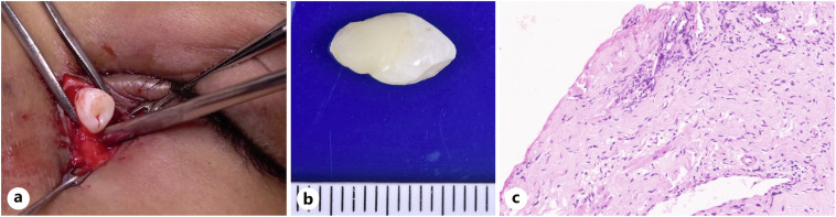
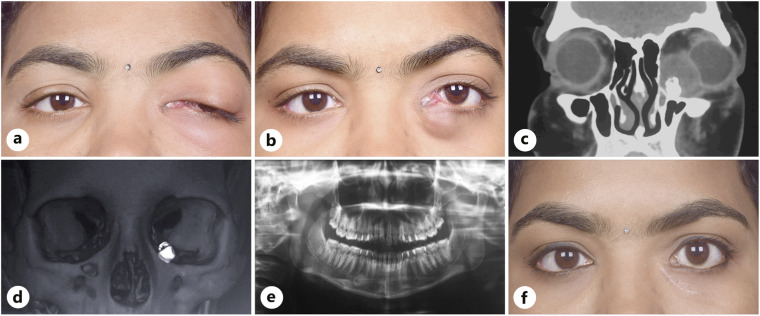
Case presentation: A 21-year-old lady presented with left inferomedial swelling, associated with pain and redness of 1-week-duration. A lacrimal sac diverticulitis with preseptal cellulitis was suspected and imaging was requested in view of progression. Computed tomography scan of orbit revealed a well-defined heterogenous mass, with associated hyperdense lesion within the mass, prompting a possibility of a foreign body granuloma. Surgical exploration and excision confirmed the diagnosis of an ectopic canine tooth with associated DC along with excessive adhesion of the cyst to the lacrimal sac wall. Maxillary sinus did not show any abnormality. Lacrimal irrigation was patent with no recurrence noted at 6-month follow-up.
Conclusion: This report highlights the presentation of DC with associated ectopic canine tooth as an isolated orbital mass, initially manifesting as preseptal cellulitis with lacrimal sac diverticulitis. Recurrent episodes of infection can lead to synechiae formation, rendering adjacent lacrimal sac more prone to injury. Cyst excision with meticulous dissection yields good outcome.
期刊介绍:
This peer-reviewed online-only journal publishes original case reports covering the entire spectrum of ophthalmology, including prevention, diagnosis, treatment, toxicities of therapy, supportive care, quality-of-life, and survivorship issues. The submission of negative results is strongly encouraged. The journal will also accept case reports dealing with the use of novel technologies, both in the arena of diagnosis and treatment. Supplementary material is welcomed. The intent of the journal is to provide clinicians and researchers with a tool to disseminate their personal experiences to a wider public as well as to review interesting cases encountered by colleagues all over the world. Universally used terms can be searched across the entire growing collection of case reports, further facilitating the retrieval of specific information. Following the open access principle, the entire contents can be retrieved at no charge, guaranteeing easy access to this valuable source of anecdotal information at all times.

 求助内容:
求助内容: 应助结果提醒方式:


