Eva Magdalene Weinberger, Loka Thangamathesvaran, J Fernando Arevalo
{"title":"Atypical COVID-19-Related Acute Macular Neuroretinopathy with Progression Resulting in Severe Vision Loss.","authors":"Eva Magdalene Weinberger, Loka Thangamathesvaran, J Fernando Arevalo","doi":"10.1159/000542802","DOIUrl":null,"url":null,"abstract":"<p><strong>Introduction: </strong>We review the clinical course of a patient with decreased vision in the setting of COVID-19 infection consistent with an atypical presentation of acute macular neuroretinopathy (AMN).</p><p><strong>Case presentation: </strong>A 56-year-old Caucasian woman developed a scotoma in the right eye 3 days after COVID-19 diagnosis and in her left eye on day 5. Baseline exam showed significantly reduced visual acuity bilaterally best corrected visual acuity of 20/60 in the right eye and 20/40 in the left eye from a patient-reported baseline of 20/20 in each eye. Examination of the fundus was remarkable for small flame-shaped hemorrhages in the superior arcade of both eyes. Near-infrared reflectance imaging revealed a singular wedge-shaped lesion in each eye close to the fovea and spectral domain-optical coherence tomography confirmed disruption of the photoreceptor layer and ellipsoid zones. Our leading diagnosis given the presentation was COVID-19-associated AMN. Given no evidence of a clear treatment, observation was selected. Three weeks later, visual acuity deteriorated further to 20/100 OD and 20/80 OS, with persistence of the wedge-shaped lesions. At 3-month follow-up, fundus photographs remained unremarkable; however, visual acuity had dropped further to 20/300 bilaterally, with persistence of the scotomata and outer retinal layer disruptions. 6 months later, treatment with a dexamethasone implant improved vision to 20/125 OD and 20/150 OS.</p><p><strong>Conclusion: </strong>Among COVID-19-induced AMN, our case is remarkable for the severe progression of visual impairment over 3 months of follow-up and improvement with a dexamethasone implant. Furthermore, absence of classical AMN lesions on fundus photography raises the question whether COVID-induced AMN may lead to a clinically distinct, potentially more severe picture than AMN arising from previously identified causes.</p>","PeriodicalId":9635,"journal":{"name":"Case Reports in Ophthalmology","volume":"16 1","pages":"41-47"},"PeriodicalIF":0.6000,"publicationDate":"2024-12-10","publicationTypes":"Journal Article","fieldsOfStudy":null,"isOpenAccess":false,"openAccessPdf":"https://www.ncbi.nlm.nih.gov/pmc/articles/PMC11842036/pdf/","citationCount":"0","resultStr":null,"platform":"Semanticscholar","paperid":null,"PeriodicalName":"Case Reports in Ophthalmology","FirstCategoryId":"1085","ListUrlMain":"https://doi.org/10.1159/000542802","RegionNum":0,"RegionCategory":null,"ArticlePicture":[],"TitleCN":null,"AbstractTextCN":null,"PMCID":null,"EPubDate":"2025/1/1 0:00:00","PubModel":"eCollection","JCR":"Q4","JCRName":"OPHTHALMOLOGY","Score":null,"Total":0}
引用次数: 0
Abstract
Introduction: We review the clinical course of a patient with decreased vision in the setting of COVID-19 infection consistent with an atypical presentation of acute macular neuroretinopathy (AMN).
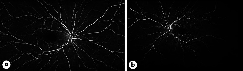
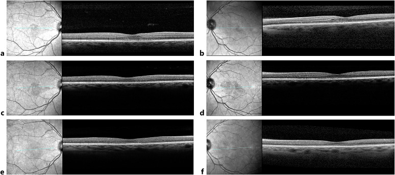
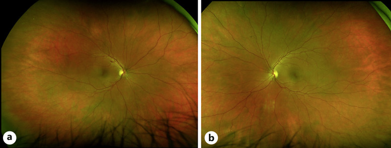
Case presentation: A 56-year-old Caucasian woman developed a scotoma in the right eye 3 days after COVID-19 diagnosis and in her left eye on day 5. Baseline exam showed significantly reduced visual acuity bilaterally best corrected visual acuity of 20/60 in the right eye and 20/40 in the left eye from a patient-reported baseline of 20/20 in each eye. Examination of the fundus was remarkable for small flame-shaped hemorrhages in the superior arcade of both eyes. Near-infrared reflectance imaging revealed a singular wedge-shaped lesion in each eye close to the fovea and spectral domain-optical coherence tomography confirmed disruption of the photoreceptor layer and ellipsoid zones. Our leading diagnosis given the presentation was COVID-19-associated AMN. Given no evidence of a clear treatment, observation was selected. Three weeks later, visual acuity deteriorated further to 20/100 OD and 20/80 OS, with persistence of the wedge-shaped lesions. At 3-month follow-up, fundus photographs remained unremarkable; however, visual acuity had dropped further to 20/300 bilaterally, with persistence of the scotomata and outer retinal layer disruptions. 6 months later, treatment with a dexamethasone implant improved vision to 20/125 OD and 20/150 OS.
Conclusion: Among COVID-19-induced AMN, our case is remarkable for the severe progression of visual impairment over 3 months of follow-up and improvement with a dexamethasone implant. Furthermore, absence of classical AMN lesions on fundus photography raises the question whether COVID-induced AMN may lead to a clinically distinct, potentially more severe picture than AMN arising from previously identified causes.
期刊介绍:
This peer-reviewed online-only journal publishes original case reports covering the entire spectrum of ophthalmology, including prevention, diagnosis, treatment, toxicities of therapy, supportive care, quality-of-life, and survivorship issues. The submission of negative results is strongly encouraged. The journal will also accept case reports dealing with the use of novel technologies, both in the arena of diagnosis and treatment. Supplementary material is welcomed. The intent of the journal is to provide clinicians and researchers with a tool to disseminate their personal experiences to a wider public as well as to review interesting cases encountered by colleagues all over the world. Universally used terms can be searched across the entire growing collection of case reports, further facilitating the retrieval of specific information. Following the open access principle, the entire contents can be retrieved at no charge, guaranteeing easy access to this valuable source of anecdotal information at all times.

 求助内容:
求助内容: 应助结果提醒方式:


