Sophie C Eberlein, Silvan Hess, Samuel F Schaible, Frank M Klenke, Andreas Hecker
{"title":"ACL tunnel placement using 3D printed surgical guides - a porcine feasibility study.","authors":"Sophie C Eberlein, Silvan Hess, Samuel F Schaible, Frank M Klenke, Andreas Hecker","doi":"10.1186/s41205-024-00215-0","DOIUrl":null,"url":null,"abstract":"<p><strong>Background: </strong>Anterior cruciate ligament reconstruction (ACLR) failures are associated with misplacement of the bone tunnels in up to 88%. The aim of this study is to evaluate the feasibility and accuracy of ACL tunnel placement performed with 3D printed guides.</p><p><strong>Methods: </strong>3D models of the femur and tibia from ten porcine specimens were reconstructed using CT scans. ACL tunnel aiming guides were created and fitted to the proximal tibial and distal femoral metaphyseal cortices. Each guide comprised two sleeves to secure the guide to the bone using Kirschner wires and one sleeve for inserting the ACL tunnel guide wire. Guides were printed using a biomedically certified resin on the in-house 3D printer. They were fixed to the antero-medial tibia/distal-lateral femur with Kirschner wires and the ACL guide wire was inserted, then the guides were removed and the ACL guide wire was drilled over. Post-operative CT scans were obtained in order to compare the actual positions of the tunnel to the planned positions. Results are presented as medians and ranges since normal distribution could not be confirmed.</p><p><strong>Result: </strong>Median deviations between preoperative plan and actual postoperative positon were 1.15 mm (0.7-3 mm) and 0.75 mm (0.3-2.8 mm) for femoral and tibial tunnels, respectively.</p><p><strong>Conclusion: </strong>Good accuracy of ACL tunnel placement can be achieved using 3D printed guides. Applied to a clinical setting, this technique has the potential to significantly reduce complications due to misplacement of bone tunnels.</p>","PeriodicalId":72036,"journal":{"name":"3D printing in medicine","volume":"11 1","pages":"6"},"PeriodicalIF":3.1000,"publicationDate":"2025-02-19","publicationTypes":"Journal Article","fieldsOfStudy":null,"isOpenAccess":false,"openAccessPdf":"https://www.ncbi.nlm.nih.gov/pmc/articles/PMC11837315/pdf/","citationCount":"0","resultStr":null,"platform":"Semanticscholar","paperid":null,"PeriodicalName":"3D printing in medicine","FirstCategoryId":"1085","ListUrlMain":"https://doi.org/10.1186/s41205-024-00215-0","RegionNum":0,"RegionCategory":null,"ArticlePicture":[],"TitleCN":null,"AbstractTextCN":null,"PMCID":null,"EPubDate":"","PubModel":"","JCR":"Q1","JCRName":"RADIOLOGY, NUCLEAR MEDICINE & MEDICAL IMAGING","Score":null,"Total":0}
引用次数: 0
Abstract
Background: Anterior cruciate ligament reconstruction (ACLR) failures are associated with misplacement of the bone tunnels in up to 88%. The aim of this study is to evaluate the feasibility and accuracy of ACL tunnel placement performed with 3D printed guides.
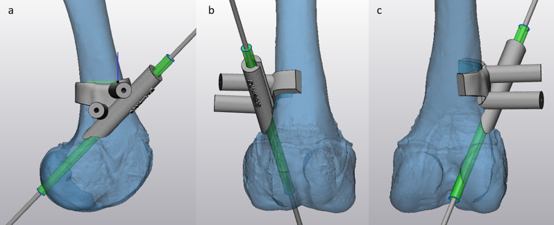
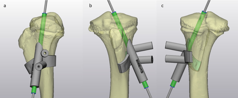
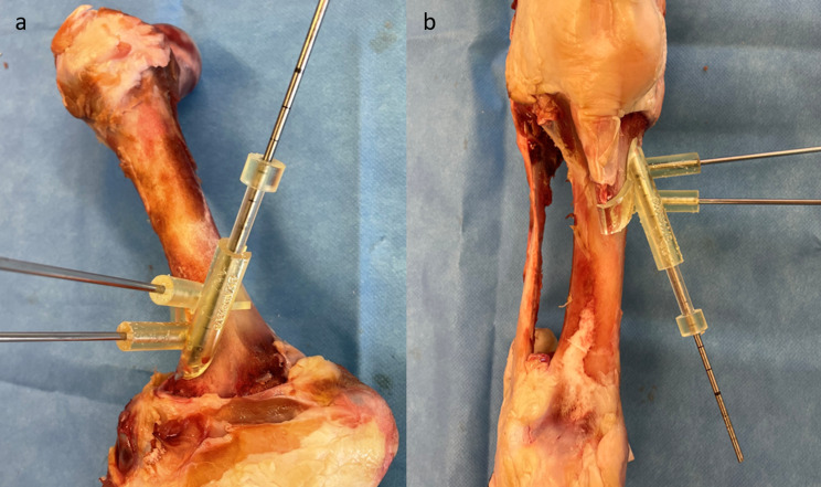
Methods: 3D models of the femur and tibia from ten porcine specimens were reconstructed using CT scans. ACL tunnel aiming guides were created and fitted to the proximal tibial and distal femoral metaphyseal cortices. Each guide comprised two sleeves to secure the guide to the bone using Kirschner wires and one sleeve for inserting the ACL tunnel guide wire. Guides were printed using a biomedically certified resin on the in-house 3D printer. They were fixed to the antero-medial tibia/distal-lateral femur with Kirschner wires and the ACL guide wire was inserted, then the guides were removed and the ACL guide wire was drilled over. Post-operative CT scans were obtained in order to compare the actual positions of the tunnel to the planned positions. Results are presented as medians and ranges since normal distribution could not be confirmed.
Result: Median deviations between preoperative plan and actual postoperative positon were 1.15 mm (0.7-3 mm) and 0.75 mm (0.3-2.8 mm) for femoral and tibial tunnels, respectively.
Conclusion: Good accuracy of ACL tunnel placement can be achieved using 3D printed guides. Applied to a clinical setting, this technique has the potential to significantly reduce complications due to misplacement of bone tunnels.
背景:高达88%的前交叉韧带重建(ACLR)失败与骨隧道错位有关。本研究的目的是评估使用3D打印导向器放置ACL隧道的可行性和准确性。方法:采用CT扫描重建10只猪的股骨和胫骨三维模型。创建ACL隧道瞄准导具并将其安装到胫骨近端和股骨远端干骺端皮质。每个导针包括两个套筒,用于用克氏针将导针固定在骨上,一个套筒用于插入ACL隧道导针。在内部3D打印机上使用生物医学认证的树脂打印指南。用克氏针将它们固定在胫骨前内侧/股骨远外侧,插入ACL导丝,然后取出导丝,钻穿ACL导丝。术后进行CT扫描以比较隧道的实际位置和计划位置。由于正态分布无法确定,因此结果以中位数和范围表示。结果:股骨和胫骨隧道的术前计划位置与术后实际位置的中位偏差分别为1.15 mm (0.7-3 mm)和0.75 mm (0.3-2.8 mm)。结论:使用3D打印支架可以获得良好的ACL隧道定位精度。应用于临床,该技术有可能显著减少由于骨隧道错位引起的并发症。

 求助内容:
求助内容: 应助结果提醒方式:


