{"title":"CT lymphangiography of the thoracic duct in mice: direct mesenteric versus popliteal lymph node puncture.","authors":"Shimpei Kato, Haruto Sugawara, Toshihiro Furuta, Osamu Abe, Hiroyuki Akai","doi":"10.1186/s41747-025-00568-z","DOIUrl":null,"url":null,"abstract":"<p><strong>Background: </strong>To evaluate the efficacy of computed tomography (CT) lymphangiography after direct mesenteric lymph node injection for thoracic duct (TD) visualization in mice.</p><p><strong>Methods: </strong>Twelve female BALB/c mice were injected with 35 μL of iodinated contrast medium (iomeprol 350 mgI/mL) into the mesenteric (mesenteric group) or popliteal (popliteal group) lymph nodes. CT images were acquired before injection and 1 min, 3 min, 5 min, 10 min, and 15 min after injection using a micro-CT scanner. Contrast ratios (CRs) were measured at the cisterna chyli and three levels of the TD (diaphragm, carina, and venous angle). Two experienced radiologists qualitatively assessed images as good, fair, or poor.</p><p><strong>Results: </strong>The mesenteric group had significantly higher mean (± standard deviation) CRs than the popliteal group for all examined regions at 1 min after injection: cisterna chyli (14.01 ± 4.77 versus 1.47 ± 1.21, p < 0.001), diaphragm (7.28 ± 2.50 versus 0.85 ± 0.61, p = 0.0011), carina (10.33 ± 3.42 versus 0.44 ± 0.40, p < 0.001), and venous angle (6.26 ± 2.02 versus 0.79 ± 0.75, p < 0.001). For the TD between the cisterna chyli and the diaphragm, 6/6 mice in the mesenteric group showed strong enhancement, whereas 5/6 mice in the popliteal group showed minimal or no enhancement. The visual scores of the mesenteric group were significantly higher than those of the popliteal group for all the evaluated regions (p = 0.002).</p><p><strong>Conclusion: </strong>CT lymphangiography via mesenteric lymph node injection provides better imaging of the TD in mice than popliteal lymph node injection.</p><p><strong>Relevance statement: </strong>This study enhances TD visualization in mice, advancing preclinical research on lymphatic disorders and improving translational applications for better clinical diagnostics and treatments.</p><p><strong>Key points: </strong>Mesenteric lymph node injection improved the efficacy of TD CT lymphangiography in mice. Mesenteric injection provided significantly better TD visualization than popliteal injection. Enhanced TD visualization in mice advances preclinical research on lymphatic diseases.</p>","PeriodicalId":36926,"journal":{"name":"European Radiology Experimental","volume":"9 1","pages":"22"},"PeriodicalIF":3.6000,"publicationDate":"2025-02-18","publicationTypes":"Journal Article","fieldsOfStudy":null,"isOpenAccess":false,"openAccessPdf":"https://www.ncbi.nlm.nih.gov/pmc/articles/PMC11836253/pdf/","citationCount":"0","resultStr":null,"platform":"Semanticscholar","paperid":null,"PeriodicalName":"European Radiology Experimental","FirstCategoryId":"1085","ListUrlMain":"https://doi.org/10.1186/s41747-025-00568-z","RegionNum":0,"RegionCategory":null,"ArticlePicture":[],"TitleCN":null,"AbstractTextCN":null,"PMCID":null,"EPubDate":"","PubModel":"","JCR":"Q1","JCRName":"RADIOLOGY, NUCLEAR MEDICINE & MEDICAL IMAGING","Score":null,"Total":0}
引用次数: 0
Abstract
Background: To evaluate the efficacy of computed tomography (CT) lymphangiography after direct mesenteric lymph node injection for thoracic duct (TD) visualization in mice.
Methods: Twelve female BALB/c mice were injected with 35 μL of iodinated contrast medium (iomeprol 350 mgI/mL) into the mesenteric (mesenteric group) or popliteal (popliteal group) lymph nodes. CT images were acquired before injection and 1 min, 3 min, 5 min, 10 min, and 15 min after injection using a micro-CT scanner. Contrast ratios (CRs) were measured at the cisterna chyli and three levels of the TD (diaphragm, carina, and venous angle). Two experienced radiologists qualitatively assessed images as good, fair, or poor.
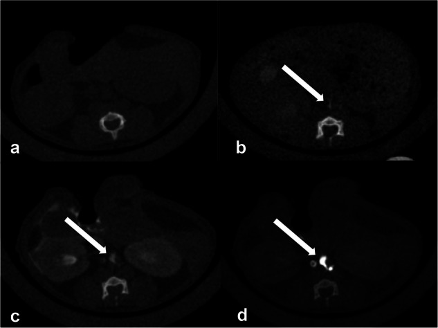
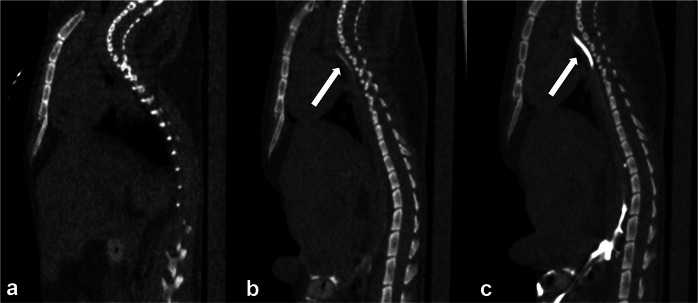
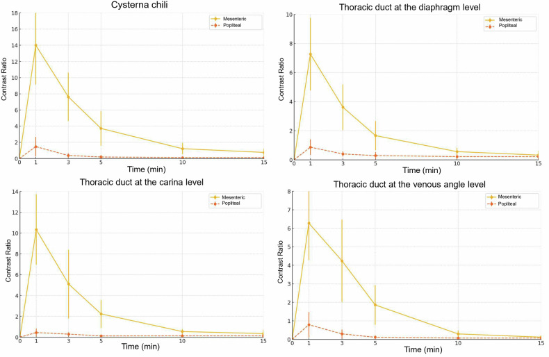
Results: The mesenteric group had significantly higher mean (± standard deviation) CRs than the popliteal group for all examined regions at 1 min after injection: cisterna chyli (14.01 ± 4.77 versus 1.47 ± 1.21, p < 0.001), diaphragm (7.28 ± 2.50 versus 0.85 ± 0.61, p = 0.0011), carina (10.33 ± 3.42 versus 0.44 ± 0.40, p < 0.001), and venous angle (6.26 ± 2.02 versus 0.79 ± 0.75, p < 0.001). For the TD between the cisterna chyli and the diaphragm, 6/6 mice in the mesenteric group showed strong enhancement, whereas 5/6 mice in the popliteal group showed minimal or no enhancement. The visual scores of the mesenteric group were significantly higher than those of the popliteal group for all the evaluated regions (p = 0.002).
Conclusion: CT lymphangiography via mesenteric lymph node injection provides better imaging of the TD in mice than popliteal lymph node injection.
Relevance statement: This study enhances TD visualization in mice, advancing preclinical research on lymphatic disorders and improving translational applications for better clinical diagnostics and treatments.
Key points: Mesenteric lymph node injection improved the efficacy of TD CT lymphangiography in mice. Mesenteric injection provided significantly better TD visualization than popliteal injection. Enhanced TD visualization in mice advances preclinical research on lymphatic diseases.

 求助内容:
求助内容: 应助结果提醒方式:


