Suha Khayri Ababneh, Ali Abu Siyam, Moath Alqaraleh, Futoon Abedrabbu Al-Rawashde, Muna M Abbas, Sokiyna Ababneh, Nihad Al-Othman, Islam Khayri Ababneh, Ahed J Alkhatib
{"title":"Exploring the Role of Ki67 in the Liver of Diabetic Rats.","authors":"Suha Khayri Ababneh, Ali Abu Siyam, Moath Alqaraleh, Futoon Abedrabbu Al-Rawashde, Muna M Abbas, Sokiyna Ababneh, Nihad Al-Othman, Islam Khayri Ababneh, Ahed J Alkhatib","doi":"10.5455/msm.2024.36.250-256","DOIUrl":null,"url":null,"abstract":"<p><strong>Background: </strong>Diabetes is not a single disease but rather, it is one aspect of metabolic syndrome. The pathologic aspects of diabetes involve cellular changes that need to be understood.</p><p><strong>Objective: </strong>The main objective of this study was to explore the role of Ki67 in the liver of diabetic rats.</p><p><strong>Methods: </strong>The study methodology involved the induction of diabetes in rats using Alloxan (120 mg/kg). A total of 20 albino rats were randomly assigned into two groups control group (N=10) and diabetes group (n=10). Diabetic group received the dose of alloxan, while the control group received similar dose of normal saline. Glucose level was monitored daily. After the end of the experiment (one -month period), all animals were terminated. Blood samples were taken to measure biochemical investigations including glucose, cholesterol, and triglycerides. Liver tissue was excised and washed with normal saline and fixed in buffered formalin (10%). Liver tissue was processed and stained by hematoxylin and eosin for routine histological examination and also stained by immunohistochemistry for Ki67 biomarker.</p><p><strong>Results: </strong>The results revealed the efficacy of the diabetic model. All biochemical investigations were significantly higher in the diabetic group compared with that of control group (p<0.001). Histological studies showed the existence of morphological alterations in cells and fatty changes in the diabetic group compared with the control group. The expression of Ki67 was significantly higher in the diabetic group compared with that in the control group (p=0.011).</p><p><strong>Conclusion: </strong>Taken together, diabetes has adverse effects on the spleen from a histological point of view, and from the expression of Ki67.</p>","PeriodicalId":94128,"journal":{"name":"Materia socio-medica","volume":"36 4","pages":"250-256"},"PeriodicalIF":0.0000,"publicationDate":"2024-01-01","publicationTypes":"Journal Article","fieldsOfStudy":null,"isOpenAccess":false,"openAccessPdf":"https://www.ncbi.nlm.nih.gov/pmc/articles/PMC11830229/pdf/","citationCount":"0","resultStr":null,"platform":"Semanticscholar","paperid":null,"PeriodicalName":"Materia socio-medica","FirstCategoryId":"1085","ListUrlMain":"https://doi.org/10.5455/msm.2024.36.250-256","RegionNum":0,"RegionCategory":null,"ArticlePicture":[],"TitleCN":null,"AbstractTextCN":null,"PMCID":null,"EPubDate":"","PubModel":"","JCR":"","JCRName":"","Score":null,"Total":0}
引用次数: 0
Abstract
Background: Diabetes is not a single disease but rather, it is one aspect of metabolic syndrome. The pathologic aspects of diabetes involve cellular changes that need to be understood.
Objective: The main objective of this study was to explore the role of Ki67 in the liver of diabetic rats.
Methods: The study methodology involved the induction of diabetes in rats using Alloxan (120 mg/kg). A total of 20 albino rats were randomly assigned into two groups control group (N=10) and diabetes group (n=10). Diabetic group received the dose of alloxan, while the control group received similar dose of normal saline. Glucose level was monitored daily. After the end of the experiment (one -month period), all animals were terminated. Blood samples were taken to measure biochemical investigations including glucose, cholesterol, and triglycerides. Liver tissue was excised and washed with normal saline and fixed in buffered formalin (10%). Liver tissue was processed and stained by hematoxylin and eosin for routine histological examination and also stained by immunohistochemistry for Ki67 biomarker.
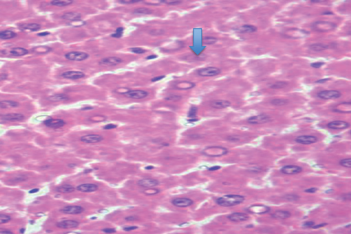
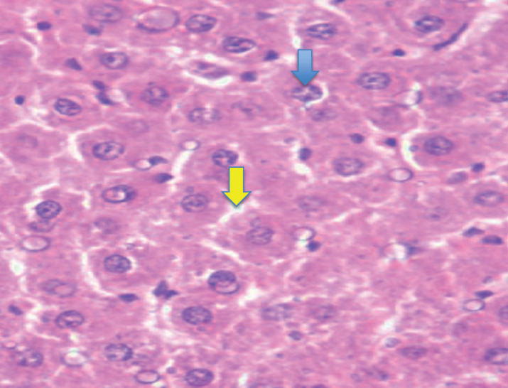
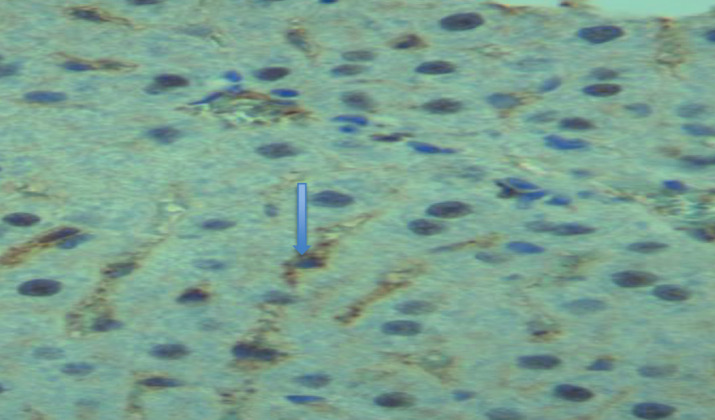
Results: The results revealed the efficacy of the diabetic model. All biochemical investigations were significantly higher in the diabetic group compared with that of control group (p<0.001). Histological studies showed the existence of morphological alterations in cells and fatty changes in the diabetic group compared with the control group. The expression of Ki67 was significantly higher in the diabetic group compared with that in the control group (p=0.011).
Conclusion: Taken together, diabetes has adverse effects on the spleen from a histological point of view, and from the expression of Ki67.

 求助内容:
求助内容: 应助结果提醒方式:


