Seung Woo Hong, Phoung Duy Dao, Kyung Won Chang, Hyun Ho Jung, Jin Woo Chang
{"title":"Minimizing Hemorrhage Complications in Deep Brain Stimulation Surgery - The Impact of Imaging Modalities and Trajectory Planning.","authors":"Seung Woo Hong, Phoung Duy Dao, Kyung Won Chang, Hyun Ho Jung, Jin Woo Chang","doi":"10.3340/jkns.2024.0198","DOIUrl":null,"url":null,"abstract":"<p><strong>Objective: </strong>This retrospective study aims to analyze hemorrhage complications in patients undergoing deep brain stimulation (DBS) surgery, focusing on the impact of imaging modalities and trajectory planning.</p><p><strong>Methods: </strong>We conducted a retrospective review of patients who underwent DBS at a single institution from September 2018 to February 2023. Surgical planning data were analyzed using a combination of 1.5 Tesla (T) and 3.0 T magnetic resonance image (MRI) for trajectory planning. Trajectories were classified into four types (type 1-4) based on the proximity of vascular structures within 2 mm on preoperative MRI scans, as defined in this study. Hemorrhage presence was evaluated through postoperative computed tomography scans.</p><p><strong>Results: </strong>Out of 200 patients analyzed, type 1 trajectories (no vascular structures within 2 mm on both MRIs) accounted for 72.70% of cases with the lowest hemorrhage rate. Significant differences in hemorrhage rates were observed among the types, with higher risks associated with type 4 trajectories. Additionally, significant variations in vascular structure types were noted across DBS targets, with subthalamic nucleus showing the highest risk.</p><p><strong>Conclusion: </strong>Meticulous trajectory planning using both 1.5 T and 3.0 T MRI is crucial in minimizing hemorrhagic complications in DBS. The study underscores the need for precise imaging and planning to enhance patient safety and surgical outcomes.</p>","PeriodicalId":16283,"journal":{"name":"Journal of Korean Neurosurgical Society","volume":" ","pages":"600-608"},"PeriodicalIF":1.7000,"publicationDate":"2025-09-01","publicationTypes":"Journal Article","fieldsOfStudy":null,"isOpenAccess":false,"openAccessPdf":"https://www.ncbi.nlm.nih.gov/pmc/articles/PMC12415488/pdf/","citationCount":"0","resultStr":null,"platform":"Semanticscholar","paperid":null,"PeriodicalName":"Journal of Korean Neurosurgical Society","FirstCategoryId":"3","ListUrlMain":"https://doi.org/10.3340/jkns.2024.0198","RegionNum":4,"RegionCategory":"医学","ArticlePicture":[],"TitleCN":null,"AbstractTextCN":null,"PMCID":null,"EPubDate":"2025/2/17 0:00:00","PubModel":"Epub","JCR":"Q4","JCRName":"CLINICAL NEUROLOGY","Score":null,"Total":0}
引用次数: 0
Abstract
Objective: This retrospective study aims to analyze hemorrhage complications in patients undergoing deep brain stimulation (DBS) surgery, focusing on the impact of imaging modalities and trajectory planning.
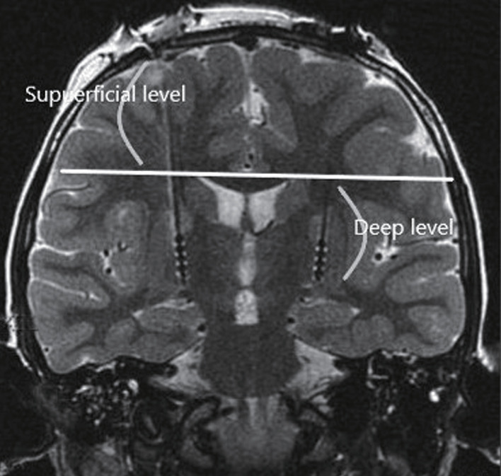
Methods: We conducted a retrospective review of patients who underwent DBS at a single institution from September 2018 to February 2023. Surgical planning data were analyzed using a combination of 1.5 Tesla (T) and 3.0 T magnetic resonance image (MRI) for trajectory planning. Trajectories were classified into four types (type 1-4) based on the proximity of vascular structures within 2 mm on preoperative MRI scans, as defined in this study. Hemorrhage presence was evaluated through postoperative computed tomography scans.
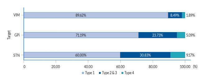
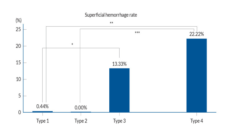
Results: Out of 200 patients analyzed, type 1 trajectories (no vascular structures within 2 mm on both MRIs) accounted for 72.70% of cases with the lowest hemorrhage rate. Significant differences in hemorrhage rates were observed among the types, with higher risks associated with type 4 trajectories. Additionally, significant variations in vascular structure types were noted across DBS targets, with subthalamic nucleus showing the highest risk.
Conclusion: Meticulous trajectory planning using both 1.5 T and 3.0 T MRI is crucial in minimizing hemorrhagic complications in DBS. The study underscores the need for precise imaging and planning to enhance patient safety and surgical outcomes.
期刊介绍:
The Journal of Korean Neurosurgical Society (J Korean Neurosurg Soc) is the official journal of the Korean Neurosurgical Society, and published bimonthly (1st day of January, March, May, July, September, and November). It launched in October 31, 1972 with Volume 1 and Number 1. J Korean Neurosurg Soc aims to allow neurosurgeons from around the world to enrich their knowledge of patient management, education, and clinical or experimental research, and hence their professionalism. This journal publishes Laboratory Investigations, Clinical Articles, Review Articles, Case Reports, Technical Notes, and Letters to the Editor. Our field of interest involves clinical neurosurgery (cerebrovascular disease, neuro-oncology, skull base neurosurgery, spine, pediatric neurosurgery, functional neurosurgery, epilepsy, neuro-trauma, and peripheral nerve disease) and laboratory work in neuroscience.
 求助内容:
求助内容: 应助结果提醒方式:


