{"title":"Quantification of Pseudomonas aeruginosa biofilms using electrochemical methods.","authors":"Lily Riordan, Perrine Lasserre, Damion Corrigan, Katherine Duncan","doi":"10.1099/acmi.0.000906.v4","DOIUrl":null,"url":null,"abstract":"<p><p>Currently, 2.29% of deaths worldwide are caused by antimicrobial resistance (AMR), compared to 1.16% from malaria and 1.55% from human immunodeficiency virus and acquired immunodeficiency syndrome. Furthermore, deaths resulting from AMR are projected to increase to more than 10 million <i>per annum</i> by 2050. Biofilms are common in hospital settings, such as medical implants, and pose a particular problem as they have shown resistance to antibiotics up to 1000-fold higher than planktonic cells because of dormant states and reduced growth rates. This is compounded by the fact that many antibiotics target mechanisms of active metabolism and are therefore less effective. The work presented here aimed to develop a method for biofilm quantification, which could be translated into the clinical setting, as well as used in the screening of antibiofilm agents. This was carried out alongside crystal violet staining, as a published point of reference. This work builds upon work previously presented by Dunphy <i>et al.</i>, in which the authors attempted to quantify the biofilm formation of <i>Pseudomonas aeruginosa</i> strain using hyperspectral imaging. Here, using electrochemical impedance spectroscopy and square wave voltammetry, the biofilm formation of two <i>P. aeruginosa</i> strains was detected within an hour after seeding <i>P. aeruginosa</i> on the sensor. A 40% decrease in impedance modulus was shown when <i>P. aeruginosa</i> biofilm had formed, compared to the media-only control. As such, this work offers a starting point for the development of real-time biofilm sensing technologies, which can be translated into implantable materials.</p>","PeriodicalId":94366,"journal":{"name":"Access microbiology","volume":"7 2","pages":""},"PeriodicalIF":0.0000,"publicationDate":"2025-02-14","publicationTypes":"Journal Article","fieldsOfStudy":null,"isOpenAccess":false,"openAccessPdf":"https://www.ncbi.nlm.nih.gov/pmc/articles/PMC11829079/pdf/","citationCount":"0","resultStr":null,"platform":"Semanticscholar","paperid":null,"PeriodicalName":"Access microbiology","FirstCategoryId":"1085","ListUrlMain":"https://doi.org/10.1099/acmi.0.000906.v4","RegionNum":0,"RegionCategory":null,"ArticlePicture":[],"TitleCN":null,"AbstractTextCN":null,"PMCID":null,"EPubDate":"2025/1/1 0:00:00","PubModel":"eCollection","JCR":"","JCRName":"","Score":null,"Total":0}
引用次数: 0
Abstract
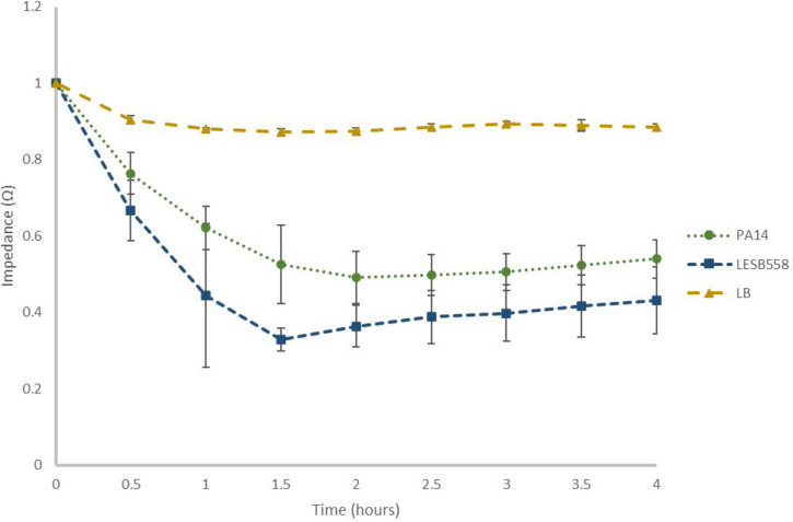
Currently, 2.29% of deaths worldwide are caused by antimicrobial resistance (AMR), compared to 1.16% from malaria and 1.55% from human immunodeficiency virus and acquired immunodeficiency syndrome. Furthermore, deaths resulting from AMR are projected to increase to more than 10 million per annum by 2050. Biofilms are common in hospital settings, such as medical implants, and pose a particular problem as they have shown resistance to antibiotics up to 1000-fold higher than planktonic cells because of dormant states and reduced growth rates. This is compounded by the fact that many antibiotics target mechanisms of active metabolism and are therefore less effective. The work presented here aimed to develop a method for biofilm quantification, which could be translated into the clinical setting, as well as used in the screening of antibiofilm agents. This was carried out alongside crystal violet staining, as a published point of reference. This work builds upon work previously presented by Dunphy et al., in which the authors attempted to quantify the biofilm formation of Pseudomonas aeruginosa strain using hyperspectral imaging. Here, using electrochemical impedance spectroscopy and square wave voltammetry, the biofilm formation of two P. aeruginosa strains was detected within an hour after seeding P. aeruginosa on the sensor. A 40% decrease in impedance modulus was shown when P. aeruginosa biofilm had formed, compared to the media-only control. As such, this work offers a starting point for the development of real-time biofilm sensing technologies, which can be translated into implantable materials.

 求助内容:
求助内容: 应助结果提醒方式:


