Maximilien Lépine, Sarah Schmitz, Svenja Körber, Kernt Köhler
{"title":"Feline malignant lymphoma in an uncommon location as a differential diagnosis for neurological disease.","authors":"Maximilien Lépine, Sarah Schmitz, Svenja Körber, Kernt Köhler","doi":"10.1177/20551169241300815","DOIUrl":null,"url":null,"abstract":"<p><strong>Case summary: </strong>A 12-year-old male castrated domestic shorthair cat exhibited right Horner's syndrome, right facial nerve paresis, difficulty swallowing, coughing, gait abnormalities and weight loss. Despite prior unspecific treatment by a primary care veterinarian with cortisone and antibiotics, the cat's condition worsened, culminating in tetraparesis and right hemispasms. Imaging studies, including CT and MRI, identified a mass extending from the carotid body into the neurocranium, causing displacement of adjacent brain structures and meningeal contrast uptake. Histopathology confirmed a malignant B-cell lymphoma. Differential diagnoses are explored, with a particular focus on carotid body tumours, which originate from the chief cells of the carotid body. These neoplasias are rare in non-human primates, dogs, cats and horses, possibly influenced by genetic predisposition and environmental factors such as hypoxia.</p><p><strong>Relevance and novel information: </strong>Carotid body tumours are rare in cats, as they are in other animal species. Although lymphomas are the most common feline neoplasms, to our knowledge, no previous case of a B-cell lymphoma in the carotid body has been described in the feline species to date. This case underscores the importance of considering rare and common neoplastic entities in feline patients with atypical clinical presentations and locations. Thereby highlighting the diagnostic challenges in veterinary oncology.</p>","PeriodicalId":36588,"journal":{"name":"Journal of Feline Medicine and Surgery Open Reports","volume":"11 1","pages":"20551169241300815"},"PeriodicalIF":0.7000,"publicationDate":"2025-02-15","publicationTypes":"Journal Article","fieldsOfStudy":null,"isOpenAccess":false,"openAccessPdf":"https://www.ncbi.nlm.nih.gov/pmc/articles/PMC11829305/pdf/","citationCount":"0","resultStr":null,"platform":"Semanticscholar","paperid":null,"PeriodicalName":"Journal of Feline Medicine and Surgery Open Reports","FirstCategoryId":"1085","ListUrlMain":"https://doi.org/10.1177/20551169241300815","RegionNum":0,"RegionCategory":null,"ArticlePicture":[],"TitleCN":null,"AbstractTextCN":null,"PMCID":null,"EPubDate":"2025/1/1 0:00:00","PubModel":"eCollection","JCR":"Q3","JCRName":"VETERINARY SCIENCES","Score":null,"Total":0}
引用次数: 0
Abstract
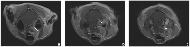
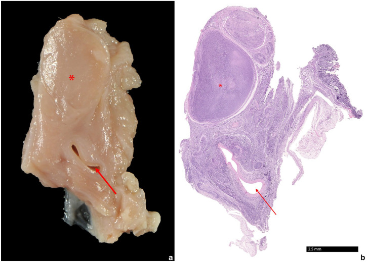
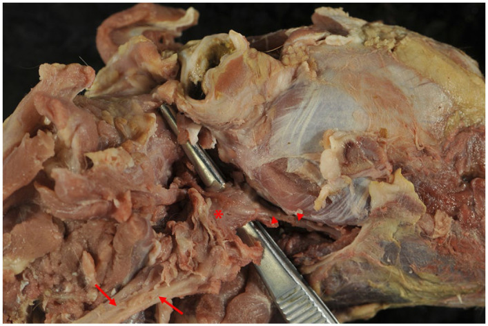
Case summary: A 12-year-old male castrated domestic shorthair cat exhibited right Horner's syndrome, right facial nerve paresis, difficulty swallowing, coughing, gait abnormalities and weight loss. Despite prior unspecific treatment by a primary care veterinarian with cortisone and antibiotics, the cat's condition worsened, culminating in tetraparesis and right hemispasms. Imaging studies, including CT and MRI, identified a mass extending from the carotid body into the neurocranium, causing displacement of adjacent brain structures and meningeal contrast uptake. Histopathology confirmed a malignant B-cell lymphoma. Differential diagnoses are explored, with a particular focus on carotid body tumours, which originate from the chief cells of the carotid body. These neoplasias are rare in non-human primates, dogs, cats and horses, possibly influenced by genetic predisposition and environmental factors such as hypoxia.
Relevance and novel information: Carotid body tumours are rare in cats, as they are in other animal species. Although lymphomas are the most common feline neoplasms, to our knowledge, no previous case of a B-cell lymphoma in the carotid body has been described in the feline species to date. This case underscores the importance of considering rare and common neoplastic entities in feline patients with atypical clinical presentations and locations. Thereby highlighting the diagnostic challenges in veterinary oncology.

 求助内容:
求助内容: 应助结果提醒方式:


