{"title":"Applications of Artificial Intelligence in Choroid Visualization for Myopia: A Comprehensive Scoping Review.","authors":"Ali M Alhalafi","doi":"10.4103/meajo.meajo_154_24","DOIUrl":null,"url":null,"abstract":"<p><p>Numerous artificial intelligence (AI) models, including deep learning techniques, are being developed to segment choroids in optical coherence tomography (OCT) images. However, there is a need for consensus on which specific models to use, requiring further synthesis of their efficacy and role in choroid visualization in myopic patients. A systematic literature search was conducted on three main databases (PubMed, Web of Science, and Scopus) using the search terms: \"Machine learning\" OR \"Artificial Intelligence\" OR \"Deep learning\" AND \"Myopia\" AND \"Choroid\" OR \"Choroidal\" from inception to February 2024 removing duplicates. A total of 12 studies were included. The populations included myopic patients with varying degrees of myopia. The AI models applied were primarily deep learning models, including U-Net with a bidirectional Convolutional Long Short-Term Memory module, LASSO regression, Attention-based Dense U-Net network, ResNeSt101 architecture training five models, and Mask Region-Based Convolutional Neural Network. The reviewed AI models demonstrated high diagnostic accuracy, including sensitivity, specificity, and area under the curve values, in identifying and assessing myopia-related changes. Various biomarkers were assessed, such as choroidal thickness, choroidal vascularity index, choroidal vessel volume, luminal volume, and stromal volume, providing valuable insights into the structural and vascular changes associated with the condition. The integration of AI models in ophthalmological imaging represents a significant advancement in the diagnosis and management of myopia. The high diagnostic accuracy and efficiency of these models underscore their potential to revolutionize myopia care, improving patient outcomes through early detection and precise monitoring of disease progression. Future studies should focus on standardizing AI methodologies and expanding their application to broader clinical settings to fully realize their potential in ophthalmology.</p>","PeriodicalId":18740,"journal":{"name":"Middle East African Journal of Ophthalmology","volume":"30 4","pages":"189-202"},"PeriodicalIF":0.3000,"publicationDate":"2024-12-02","publicationTypes":"Journal Article","fieldsOfStudy":null,"isOpenAccess":false,"openAccessPdf":"https://www.ncbi.nlm.nih.gov/pmc/articles/PMC11823532/pdf/","citationCount":"0","resultStr":null,"platform":"Semanticscholar","paperid":null,"PeriodicalName":"Middle East African Journal of Ophthalmology","FirstCategoryId":"1085","ListUrlMain":"https://doi.org/10.4103/meajo.meajo_154_24","RegionNum":0,"RegionCategory":null,"ArticlePicture":[],"TitleCN":null,"AbstractTextCN":null,"PMCID":null,"EPubDate":"2023/10/1 0:00:00","PubModel":"eCollection","JCR":"Q4","JCRName":"OPHTHALMOLOGY","Score":null,"Total":0}
引用次数: 0
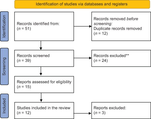
Abstract
Numerous artificial intelligence (AI) models, including deep learning techniques, are being developed to segment choroids in optical coherence tomography (OCT) images. However, there is a need for consensus on which specific models to use, requiring further synthesis of their efficacy and role in choroid visualization in myopic patients. A systematic literature search was conducted on three main databases (PubMed, Web of Science, and Scopus) using the search terms: "Machine learning" OR "Artificial Intelligence" OR "Deep learning" AND "Myopia" AND "Choroid" OR "Choroidal" from inception to February 2024 removing duplicates. A total of 12 studies were included. The populations included myopic patients with varying degrees of myopia. The AI models applied were primarily deep learning models, including U-Net with a bidirectional Convolutional Long Short-Term Memory module, LASSO regression, Attention-based Dense U-Net network, ResNeSt101 architecture training five models, and Mask Region-Based Convolutional Neural Network. The reviewed AI models demonstrated high diagnostic accuracy, including sensitivity, specificity, and area under the curve values, in identifying and assessing myopia-related changes. Various biomarkers were assessed, such as choroidal thickness, choroidal vascularity index, choroidal vessel volume, luminal volume, and stromal volume, providing valuable insights into the structural and vascular changes associated with the condition. The integration of AI models in ophthalmological imaging represents a significant advancement in the diagnosis and management of myopia. The high diagnostic accuracy and efficiency of these models underscore their potential to revolutionize myopia care, improving patient outcomes through early detection and precise monitoring of disease progression. Future studies should focus on standardizing AI methodologies and expanding their application to broader clinical settings to fully realize their potential in ophthalmology.
许多人工智能(AI)模型,包括深度学习技术,正在开发用于分割光学相干断层扫描(OCT)图像中的脉络膜。然而,需要就具体使用哪种模型达成共识,需要进一步综合它们在近视患者脉络膜显像中的疗效和作用。在三个主要数据库(PubMed、Web of Science和Scopus)上进行了系统的文献检索,检索词为:“机器学习”或“人工智能”或“深度学习”和“近视”和“Choroid”或“脉络膜”,检索时间从成立到2024年2月。共纳入12项研究。人群包括不同程度的近视患者。应用的人工智能模型主要是深度学习模型,包括带有双向卷积长短期记忆模块的U-Net、LASSO回归、基于注意力的密集U-Net网络、resnesst101架构训练的五个模型以及基于Mask区域的卷积神经网络。所回顾的人工智能模型在识别和评估近视相关变化方面表现出很高的诊断准确性,包括敏感性、特异性和曲线下面积值。评估了各种生物标志物,如脉络膜厚度、脉络膜血管指数、脉络膜血管体积、管腔体积和间质体积,为与该疾病相关的结构和血管变化提供了有价值的见解。人工智能模型在眼科成像中的集成代表了近视诊断和治疗的重大进步。这些模型的高诊断准确性和效率强调了它们在近视护理方面的潜力,通过早期发现和精确监测疾病进展来改善患者的预后。未来的研究应着眼于规范人工智能方法,并将其应用到更广泛的临床环境中,以充分发挥其在眼科中的潜力。
期刊介绍:
The Middle East African Journal of Ophthalmology (MEAJO), published four times per year in print and online, is an official journal of the Middle East African Council of Ophthalmology (MEACO). It is an international, peer-reviewed journal whose mission includes publication of original research of interest to ophthalmologists in the Middle East and Africa, and to provide readers with high quality educational review articles from world-renown experts. MEAJO, previously known as Middle East Journal of Ophthalmology (MEJO) was founded by Dr Akef El Maghraby in 1993.

 求助内容:
求助内容: 应助结果提醒方式:


