Dimensional changes in buccal cortical bone and lesion volume in teeth with persistent chronic periapical disease subjected to periapical surgery: a cone beam computed tomography study at one year of follow-up.
A Boronat-López, J-C Bernabeu-Mira, M Peñarrocha-Diago, M Peñarrocha-Diago, D Peñarrocha-Oltra
{"title":"Dimensional changes in buccal cortical bone and lesion volume in teeth with persistent chronic periapical disease subjected to periapical surgery: a cone beam computed tomography study at one year of follow-up.","authors":"A Boronat-López, J-C Bernabeu-Mira, M Peñarrocha-Diago, M Peñarrocha-Diago, D Peñarrocha-Oltra","doi":"10.4317/medoral.27006","DOIUrl":null,"url":null,"abstract":"<p><strong>Background: </strong>This study aimed to evaluate changes in buccal cortical bone and lesion volume in teeth with persistent periapical disease one year after periapical surgery using cone-beam computed tomography (CBCT).</p><p><strong>Material and methods: </strong>A prospective study was conducted involving patients with persistent periapical disease undergoing periapical surgery, with one year of follow-up. Data collected included patient age, gender, teeth involved, and the number of roots/lesions. CBCT measurements were taken preoperatively and one year post-surgery, including the distance from the cementoenamel junction to the buccal bone crest (CEJ-BBC), marginal bone loss, buccal cortical height, presence of fenestration, apical depth, cortical bone width at 1, 3, and 5 mm from the buccal bone crest, and lesion volume in mm³. Success was assessed using the \"Modified Penn 3D criteria.\"</p><p><strong>Results: </strong>The study included 92 patients with 111 roots exhibiting persistent chronic periapical lesions. Statistically significant changes were observed in all buccal cortical bone parameters one year after surgery. The CEJ-BBC distance increased, indicating a marginal bone loss of 0.23 mm. Notably, the height from the buccal cortical bone crest to the lesion, apical depth, buccal bone thickness, the number of fenestrations, and lesion volume decreased (91.1%). Buccal cortical bone thickness was a predictor of volume reduction, showing a significant relationship at T1 between greater thickness and smaller volume variation. Patient age and gender did not significantly influence these changes. Fenestrations and larger lesion volumes correlated with reduced healing probabilities. The overall success rate was 88%, with tooth position and root involvement impacting healing outcomes.</p><p><strong>Conclusions: </strong>One year post-surgery, buccal cortical bone showed no clinically relevant changes, while lesion volume decreased by 91.1%, more significantly in anterior teeth. Greater buccal cortical bone width was associated with smaller volume reduction. A larger lesion volume and presence of fenestrations adversely affected healing rates.</p>","PeriodicalId":49016,"journal":{"name":"Medicina Oral Patologia Oral Y Cirugia Bucal","volume":" ","pages":"e469-e475"},"PeriodicalIF":2.1000,"publicationDate":"2025-05-01","publicationTypes":"Journal Article","fieldsOfStudy":null,"isOpenAccess":false,"openAccessPdf":"https://www.ncbi.nlm.nih.gov/pmc/articles/PMC12019659/pdf/","citationCount":"0","resultStr":null,"platform":"Semanticscholar","paperid":null,"PeriodicalName":"Medicina Oral Patologia Oral Y Cirugia Bucal","FirstCategoryId":"3","ListUrlMain":"https://doi.org/10.4317/medoral.27006","RegionNum":3,"RegionCategory":"医学","ArticlePicture":[],"TitleCN":null,"AbstractTextCN":null,"PMCID":null,"EPubDate":"","PubModel":"","JCR":"Q2","JCRName":"DENTISTRY, ORAL SURGERY & MEDICINE","Score":null,"Total":0}
引用次数: 0
Abstract
Background: This study aimed to evaluate changes in buccal cortical bone and lesion volume in teeth with persistent periapical disease one year after periapical surgery using cone-beam computed tomography (CBCT).
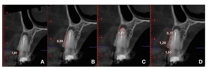
Material and methods: A prospective study was conducted involving patients with persistent periapical disease undergoing periapical surgery, with one year of follow-up. Data collected included patient age, gender, teeth involved, and the number of roots/lesions. CBCT measurements were taken preoperatively and one year post-surgery, including the distance from the cementoenamel junction to the buccal bone crest (CEJ-BBC), marginal bone loss, buccal cortical height, presence of fenestration, apical depth, cortical bone width at 1, 3, and 5 mm from the buccal bone crest, and lesion volume in mm³. Success was assessed using the "Modified Penn 3D criteria."
Results: The study included 92 patients with 111 roots exhibiting persistent chronic periapical lesions. Statistically significant changes were observed in all buccal cortical bone parameters one year after surgery. The CEJ-BBC distance increased, indicating a marginal bone loss of 0.23 mm. Notably, the height from the buccal cortical bone crest to the lesion, apical depth, buccal bone thickness, the number of fenestrations, and lesion volume decreased (91.1%). Buccal cortical bone thickness was a predictor of volume reduction, showing a significant relationship at T1 between greater thickness and smaller volume variation. Patient age and gender did not significantly influence these changes. Fenestrations and larger lesion volumes correlated with reduced healing probabilities. The overall success rate was 88%, with tooth position and root involvement impacting healing outcomes.
Conclusions: One year post-surgery, buccal cortical bone showed no clinically relevant changes, while lesion volume decreased by 91.1%, more significantly in anterior teeth. Greater buccal cortical bone width was associated with smaller volume reduction. A larger lesion volume and presence of fenestrations adversely affected healing rates.
期刊介绍:
1. Oral Medicine and Pathology:
Clinicopathological as well as medical or surgical management aspects of
diseases affecting oral mucosa, salivary glands, maxillary bones, as well as
orofacial neurological disorders, and systemic conditions with an impact on
the oral cavity.
2. Oral Surgery:
Surgical management aspects of diseases affecting oral mucosa, salivary glands,
maxillary bones, teeth, implants, oral surgical procedures. Surgical management
of diseases affecting head and neck areas.
3. Medically compromised patients in Dentistry:
Articles discussing medical problems in Odontology will also be included, with
a special focus on the clinico-odontological management of medically compromised patients, and considerations regarding high-risk or disabled patients.
4. Implantology
5. Periodontology

 求助内容:
求助内容: 应助结果提醒方式:


