Xia Li, Feng Qiao, Jiansheng Guo, Ting Jiang, Huifang Lou, Huixia Li, Gangcai Xie, Hangjun Wu, Weizhen Wang, Ruoyu Pei, Sha Liu, Mei Ye, Jin Li, Shiqin Huang, Mengya Zhang, Chaoye Ma, Yiwen Huang, Shushu Xu, Xiaofeng Li, Xiao Sun, Jun Yu, Kin Lam Fok, Shumin Duan, Hao Chen
{"title":"In situ architecture of the intercellular organelle reservoir between epididymal epithelial cells by volume electron microscopy","authors":"Xia Li, Feng Qiao, Jiansheng Guo, Ting Jiang, Huifang Lou, Huixia Li, Gangcai Xie, Hangjun Wu, Weizhen Wang, Ruoyu Pei, Sha Liu, Mei Ye, Jin Li, Shiqin Huang, Mengya Zhang, Chaoye Ma, Yiwen Huang, Shushu Xu, Xiaofeng Li, Xiao Sun, Jun Yu, Kin Lam Fok, Shumin Duan, Hao Chen","doi":"10.1038/s41467-025-56807-9","DOIUrl":null,"url":null,"abstract":"<p>Mammalian epididymal epithelial cells are crucial for sperm maturation. Historically, vacuole-like ultrastructures in epididymal epithelial cells were observed via transmission electron microscopy but were undefined. Here, we utilize volume electron microscopy (vEM) to generate 3D reconstructions of epididymal epithelial cells and identify these vacuoles as intercellular organelle reservoirs (IORs) in the lateral intercellular space (LIS), which contains protein aggregates, autophagosomes, lysosome-related organelles and mitochondrial residues. Immunolabelling of organelle markers such as P62, LC3, LAMP1 and TOMM20 confirm these findings. The IOR size or number varies across four epididymal regions and decreases with age. Rab27a mutant mice exhibit reduced IORs in the caput epididymis and a subfertility phenotype, suggesting the involvement of Rab27a in the formation of IORs. Furthermore, we observe the presence of IORs between intestinal epithelial cells besides epididymis. Amino acid transporters at IOR edges suggest dynamic protein recycling. Our findings reveal that the IOR is an important structure critical for organelle turnover and recycling outside epithelial cells with limited self-degradation capabilities.</p>","PeriodicalId":19066,"journal":{"name":"Nature Communications","volume":"35 1","pages":""},"PeriodicalIF":15.7000,"publicationDate":"2025-02-15","publicationTypes":"Journal Article","fieldsOfStudy":null,"isOpenAccess":false,"openAccessPdf":"","citationCount":"0","resultStr":null,"platform":"Semanticscholar","paperid":null,"PeriodicalName":"Nature Communications","FirstCategoryId":"103","ListUrlMain":"https://doi.org/10.1038/s41467-025-56807-9","RegionNum":1,"RegionCategory":"综合性期刊","ArticlePicture":[],"TitleCN":null,"AbstractTextCN":null,"PMCID":null,"EPubDate":"","PubModel":"","JCR":"Q1","JCRName":"MULTIDISCIPLINARY SCIENCES","Score":null,"Total":0}
引用次数: 0
Abstract
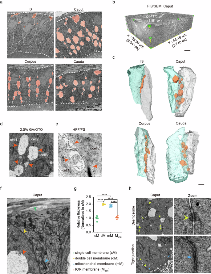
Mammalian epididymal epithelial cells are crucial for sperm maturation. Historically, vacuole-like ultrastructures in epididymal epithelial cells were observed via transmission electron microscopy but were undefined. Here, we utilize volume electron microscopy (vEM) to generate 3D reconstructions of epididymal epithelial cells and identify these vacuoles as intercellular organelle reservoirs (IORs) in the lateral intercellular space (LIS), which contains protein aggregates, autophagosomes, lysosome-related organelles and mitochondrial residues. Immunolabelling of organelle markers such as P62, LC3, LAMP1 and TOMM20 confirm these findings. The IOR size or number varies across four epididymal regions and decreases with age. Rab27a mutant mice exhibit reduced IORs in the caput epididymis and a subfertility phenotype, suggesting the involvement of Rab27a in the formation of IORs. Furthermore, we observe the presence of IORs between intestinal epithelial cells besides epididymis. Amino acid transporters at IOR edges suggest dynamic protein recycling. Our findings reveal that the IOR is an important structure critical for organelle turnover and recycling outside epithelial cells with limited self-degradation capabilities.
期刊介绍:
Nature Communications, an open-access journal, publishes high-quality research spanning all areas of the natural sciences. Papers featured in the journal showcase significant advances relevant to specialists in each respective field. With a 2-year impact factor of 16.6 (2022) and a median time of 8 days from submission to the first editorial decision, Nature Communications is committed to rapid dissemination of research findings. As a multidisciplinary journal, it welcomes contributions from biological, health, physical, chemical, Earth, social, mathematical, applied, and engineering sciences, aiming to highlight important breakthroughs within each domain.
 求助内容:
求助内容: 应助结果提醒方式:


