Mamata S Kamat, Uma V Datar, Sanjay S Byakodi, Amit A Basannavar
{"title":"Spindle cell variant of ameloblastic carcinoma: A diagnostic challenge.","authors":"Mamata S Kamat, Uma V Datar, Sanjay S Byakodi, Amit A Basannavar","doi":"10.4103/jomfp.jomfp_272_24","DOIUrl":null,"url":null,"abstract":"<p><p>An 18-year-old male presented with a 3-week history of progressively enlarging nodular growth in the posterior right mandibular region. The patient denied the use of tobacco in any form or history of trauma, and his family history was unremarkable. Clinical examination revealed a 2 × 3 cm reddish-pink, firm lesion with palpable submandibular lymph nodes. Radiological imaging showed a well-defined radiolucency distal to tooth #47. An incisional biopsy revealed the proliferation of spindle cells and ameloblastomatous epithelium showing features like pleomorphism, cellular crowding, and mitotic figures. Immunohistochemical analysis confirmed odontogenic origin. Based on the above findings, the diagnosis of the spindle cell variant of ameloblastic carcinoma (AC) was rendered. AC, constituting less than 2% of odontogenic tumours, and the spindle cell variant, a rare subtype with fewer than 15 reported cases, pose diagnostic challenges, necessitating careful histopathological and immunohistochemical evaluation for accurate diagnosis and treatment planning.</p>","PeriodicalId":38846,"journal":{"name":"Journal of Oral and Maxillofacial Pathology","volume":"28 4","pages":"713-715"},"PeriodicalIF":0.0000,"publicationDate":"2024-10-01","publicationTypes":"Journal Article","fieldsOfStudy":null,"isOpenAccess":false,"openAccessPdf":"https://www.ncbi.nlm.nih.gov/pmc/articles/PMC11819649/pdf/","citationCount":"0","resultStr":null,"platform":"Semanticscholar","paperid":null,"PeriodicalName":"Journal of Oral and Maxillofacial Pathology","FirstCategoryId":"1085","ListUrlMain":"https://doi.org/10.4103/jomfp.jomfp_272_24","RegionNum":0,"RegionCategory":null,"ArticlePicture":[],"TitleCN":null,"AbstractTextCN":null,"PMCID":null,"EPubDate":"2024/12/31 0:00:00","PubModel":"Epub","JCR":"Q3","JCRName":"Medicine","Score":null,"Total":0}
引用次数: 0
Abstract
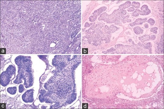
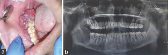
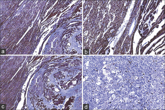
An 18-year-old male presented with a 3-week history of progressively enlarging nodular growth in the posterior right mandibular region. The patient denied the use of tobacco in any form or history of trauma, and his family history was unremarkable. Clinical examination revealed a 2 × 3 cm reddish-pink, firm lesion with palpable submandibular lymph nodes. Radiological imaging showed a well-defined radiolucency distal to tooth #47. An incisional biopsy revealed the proliferation of spindle cells and ameloblastomatous epithelium showing features like pleomorphism, cellular crowding, and mitotic figures. Immunohistochemical analysis confirmed odontogenic origin. Based on the above findings, the diagnosis of the spindle cell variant of ameloblastic carcinoma (AC) was rendered. AC, constituting less than 2% of odontogenic tumours, and the spindle cell variant, a rare subtype with fewer than 15 reported cases, pose diagnostic challenges, necessitating careful histopathological and immunohistochemical evaluation for accurate diagnosis and treatment planning.
期刊介绍:
The journal of Oral and Maxillofacial Pathology [ISSN:print-(0973-029X, online-1998-393X)] is a tri-annual journal published on behalf of “The Indian Association of Oral and Maxillofacial Pathologists” (IAOMP). The publication of JOMFP was started in the year 1993. The journal publishes papers on a wide spectrum of topics associated with the scope of Oral and Maxillofacial Pathology, also, ensuring scientific merit and quality. It is a comprehensive reading material for the professionals who want to upgrade their diagnostic skills in Oral Diseases; allows exposure to newer topics and methods of research in the Oral-facial Tissues and Pathology. New features allow an open minded thinking and approach to various pathologies. It also encourages authors to showcase quality work done by them and to compile relevant cases which are diagnostically challenging. The Journal takes pride in maintaining the quality of articles and photomicrographs.

 求助内容:
求助内容: 应助结果提醒方式:


