Manti Vijayalakshmi, Bandela Rajasekhar, Vishwaprakash Shetty, Animelli Jacob Prakash, Thokala Madhusudhan Rao, H Aparna Latha
{"title":"Histomorphometric analysis of lichen planus and lichenoid lesions - Retrospective study.","authors":"Manti Vijayalakshmi, Bandela Rajasekhar, Vishwaprakash Shetty, Animelli Jacob Prakash, Thokala Madhusudhan Rao, H Aparna Latha","doi":"10.4103/jomfp.jomfp_29_24","DOIUrl":null,"url":null,"abstract":"<p><strong>Introduction: </strong>Though it has a different aetiology, lichenoid lesions and lichen planus are both chronic inflammatory mucocutaneous conditions with similar clinical and histological characteristics. Uncertainty persists despite the development of recommendations for distinguishing oral lichen planus from lichenoid diseases.</p><p><strong>Materials and methodology: </strong>A retrospective analysis was conducted on 25 cases of oral lichen planus (OLP), oral lichenoid lesions (OLL), and normal oral mucosa (NOM) that had been histologically diagnosed. Using imagej analysis software, morphometric assessments of the cellular area, nuclear area, nucleus cytoplasmic ratio, nuclear diameter, and cellular diameter of suprabasal cells were performed on sections stained with haematoxylin and eosin. The data were then analysed using ANOVA.</p><p><strong>Results: </strong>Our study's findings demonstrated that, in comparison to the normal oral mucosa, the mean values of the nuclear area, cellular area, nuclear diameter, and cellular diameter in oral lichen planus and oral lichenoid lesions were higher.</p><p><strong>Conclusion: </strong>In the current investigation, we also found that, in comparison to normal mucosa, the nuclear-cytoplasmic ratio was lower in oral lichen planus and oral lichenoid lesions.</p>","PeriodicalId":38846,"journal":{"name":"Journal of Oral and Maxillofacial Pathology","volume":"28 4","pages":"570-575"},"PeriodicalIF":0.0000,"publicationDate":"2024-10-01","publicationTypes":"Journal Article","fieldsOfStudy":null,"isOpenAccess":false,"openAccessPdf":"https://www.ncbi.nlm.nih.gov/pmc/articles/PMC11819632/pdf/","citationCount":"0","resultStr":null,"platform":"Semanticscholar","paperid":null,"PeriodicalName":"Journal of Oral and Maxillofacial Pathology","FirstCategoryId":"1085","ListUrlMain":"https://doi.org/10.4103/jomfp.jomfp_29_24","RegionNum":0,"RegionCategory":null,"ArticlePicture":[],"TitleCN":null,"AbstractTextCN":null,"PMCID":null,"EPubDate":"2024/12/31 0:00:00","PubModel":"Epub","JCR":"Q3","JCRName":"Medicine","Score":null,"Total":0}
引用次数: 0
Abstract
Introduction: Though it has a different aetiology, lichenoid lesions and lichen planus are both chronic inflammatory mucocutaneous conditions with similar clinical and histological characteristics. Uncertainty persists despite the development of recommendations for distinguishing oral lichen planus from lichenoid diseases.
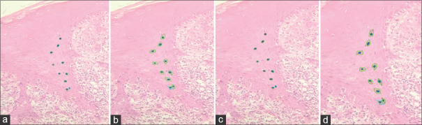
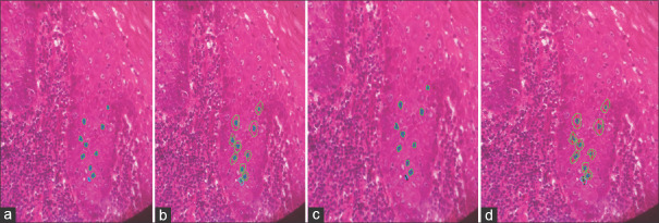
Materials and methodology: A retrospective analysis was conducted on 25 cases of oral lichen planus (OLP), oral lichenoid lesions (OLL), and normal oral mucosa (NOM) that had been histologically diagnosed. Using imagej analysis software, morphometric assessments of the cellular area, nuclear area, nucleus cytoplasmic ratio, nuclear diameter, and cellular diameter of suprabasal cells were performed on sections stained with haematoxylin and eosin. The data were then analysed using ANOVA.
Results: Our study's findings demonstrated that, in comparison to the normal oral mucosa, the mean values of the nuclear area, cellular area, nuclear diameter, and cellular diameter in oral lichen planus and oral lichenoid lesions were higher.
Conclusion: In the current investigation, we also found that, in comparison to normal mucosa, the nuclear-cytoplasmic ratio was lower in oral lichen planus and oral lichenoid lesions.
期刊介绍:
The journal of Oral and Maxillofacial Pathology [ISSN:print-(0973-029X, online-1998-393X)] is a tri-annual journal published on behalf of “The Indian Association of Oral and Maxillofacial Pathologists” (IAOMP). The publication of JOMFP was started in the year 1993. The journal publishes papers on a wide spectrum of topics associated with the scope of Oral and Maxillofacial Pathology, also, ensuring scientific merit and quality. It is a comprehensive reading material for the professionals who want to upgrade their diagnostic skills in Oral Diseases; allows exposure to newer topics and methods of research in the Oral-facial Tissues and Pathology. New features allow an open minded thinking and approach to various pathologies. It also encourages authors to showcase quality work done by them and to compile relevant cases which are diagnostically challenging. The Journal takes pride in maintaining the quality of articles and photomicrographs.


 求助内容:
求助内容: 应助结果提醒方式:


