{"title":"Gastric mucormycosis in a cat.","authors":"Emilio Mavilio, Enrico Bottero","doi":"10.1177/20551169241301914","DOIUrl":null,"url":null,"abstract":"<p><strong>Case summary: </strong>This report describes a case of gastric mucormycosis in a young Ragdoll cat with a 5-day history of vomiting. Physical examination detected mild dehydration and tenderness was elicited on abdominal palpation. The results of blood work-up and radiographic study were unremarkable; however, abdominal ultrasonographic examination revealed multiple hyperechoic neoformations at the level of the pyloric antrum, which were confirmed on endoscopic examination. Non-septate hyphae of irregular diameter with a branched appearance were observed on cytology, and histological examination revealed severe diffuse necrotising and granulomatous gastritis with the presence of intralesional fungal hyphae indicative of mucormycosis, which was confirmed by PCR tests. Antifungal therapy with ketoconazole in addition to supportive treatment temporarily improved the clinical condition. Lethargy, fever and abdominal effusion developed in the following days. Cytological examination of abdominal fluid was compatible with septic peritonitis and, given the severity of the condition, euthanasia was opted by the owners. Post-mortem examination confirmed septic peritonitis resulting from perforation of the gastric wall at one of the neoformations of the pyloric antrum.</p><p><strong>Relevance and novel information: </strong>To the authors' knowledge, this is the first reported case of gastric mucormycosis in a cat. Previous literature includes a case of mucormycosis in a Persian cat affecting only the duodenum. In both the Persian cat and the cat described here, gastrointestinal mucormycosis disease progressed rapidly and was fatal.</p>","PeriodicalId":36588,"journal":{"name":"Journal of Feline Medicine and Surgery Open Reports","volume":"11 1","pages":"20551169241301914"},"PeriodicalIF":0.7000,"publicationDate":"2025-02-12","publicationTypes":"Journal Article","fieldsOfStudy":null,"isOpenAccess":false,"openAccessPdf":"https://www.ncbi.nlm.nih.gov/pmc/articles/PMC11822825/pdf/","citationCount":"0","resultStr":null,"platform":"Semanticscholar","paperid":null,"PeriodicalName":"Journal of Feline Medicine and Surgery Open Reports","FirstCategoryId":"1085","ListUrlMain":"https://doi.org/10.1177/20551169241301914","RegionNum":0,"RegionCategory":null,"ArticlePicture":[],"TitleCN":null,"AbstractTextCN":null,"PMCID":null,"EPubDate":"2025/1/1 0:00:00","PubModel":"eCollection","JCR":"Q3","JCRName":"VETERINARY SCIENCES","Score":null,"Total":0}
引用次数: 0
Abstract
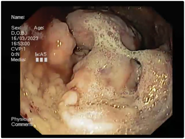
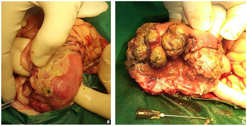
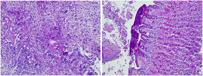
Case summary: This report describes a case of gastric mucormycosis in a young Ragdoll cat with a 5-day history of vomiting. Physical examination detected mild dehydration and tenderness was elicited on abdominal palpation. The results of blood work-up and radiographic study were unremarkable; however, abdominal ultrasonographic examination revealed multiple hyperechoic neoformations at the level of the pyloric antrum, which were confirmed on endoscopic examination. Non-septate hyphae of irregular diameter with a branched appearance were observed on cytology, and histological examination revealed severe diffuse necrotising and granulomatous gastritis with the presence of intralesional fungal hyphae indicative of mucormycosis, which was confirmed by PCR tests. Antifungal therapy with ketoconazole in addition to supportive treatment temporarily improved the clinical condition. Lethargy, fever and abdominal effusion developed in the following days. Cytological examination of abdominal fluid was compatible with septic peritonitis and, given the severity of the condition, euthanasia was opted by the owners. Post-mortem examination confirmed septic peritonitis resulting from perforation of the gastric wall at one of the neoformations of the pyloric antrum.
Relevance and novel information: To the authors' knowledge, this is the first reported case of gastric mucormycosis in a cat. Previous literature includes a case of mucormycosis in a Persian cat affecting only the duodenum. In both the Persian cat and the cat described here, gastrointestinal mucormycosis disease progressed rapidly and was fatal.

 求助内容:
求助内容: 应助结果提醒方式:


