Miguel Santos, Afonso Lima-Cabrita, Rafael Barão, Luís Abegão-Pinto
{"title":"Detection of iridocorneal angle changes after uncomplicated phacoemulsification using automated gonioscopy imaging.","authors":"Miguel Santos, Afonso Lima-Cabrita, Rafael Barão, Luís Abegão-Pinto","doi":"10.4103/sjopt.sjopt_141_24","DOIUrl":null,"url":null,"abstract":"<p><strong>Purpose: </strong>Phacoemulsification cataract surgery (PCS) is known to change anterior chamber morphology, widening the iridocorneal angle (ICA) of most patients. This study aims to determine whether automated gonioscopy imaging (AGI) with GS-1<sup>®</sup> (NIDEK CO., Japan) can detect changes in ICA morphology and pigmentation after PCS.</p><p><strong>Methods: </strong>A prospective, observational study including patients who underwent PCS at Hospital Santa Maria, Lisbon, from March to October 2022. AGI was done at the preoperative visit and 6-weeks postoperative. The images were analyzed using NAVIS-EX Software (NIDEK CO., Japan) by a masked observer. Each eye was divided into four quadrants and their morphology and pigmentation was graded according to the Shaffer and modified Scheie classifications, respectively.</p><p><strong>Results: </strong>Twenty-two eyes from 21 patients (62% female) were included, with a mean age of 74.5 ± 7.9 years. Baseline AGI identified 7 (33%) patients with angle closure. PCS led to all patients achieving open angle status and lower preoperative Shaffer grades achieved greater postoperative improvement. Statistically significant ICA widening was found in the superior (<i>P</i> = 0.004), inferior (<i>P</i> = 0.008), and temporal (<i>P</i> = 0.023) quadrants, but not the nasal quadrant (<i>P</i> = 0.21). Similarly, a statistically significant pigmentation increase was found in the superior (<i>P</i> = 0.008), inferior (<i>P</i> = 0.002), and temporal (<i>P</i> = 0.016) quadrants and less pigmented baseline quadrants showed a greater gain. The most significant pigmentation gain was in the inferior quadrant.</p><p><strong>Conclusion: </strong>Unlike other imaging modalities, AGI with GS-1 can detect changes in ICA morphology and pigmentation after routine cataract surgery, mainly in the superior, inferior, and temporal quadrants.</p>","PeriodicalId":46810,"journal":{"name":"Saudi Journal of Ophthalmology","volume":"38 4","pages":"348-351"},"PeriodicalIF":1.2000,"publicationDate":"2024-10-22","publicationTypes":"Journal Article","fieldsOfStudy":null,"isOpenAccess":false,"openAccessPdf":"https://www.ncbi.nlm.nih.gov/pmc/articles/PMC11811409/pdf/","citationCount":"0","resultStr":null,"platform":"Semanticscholar","paperid":null,"PeriodicalName":"Saudi Journal of Ophthalmology","FirstCategoryId":"1085","ListUrlMain":"https://doi.org/10.4103/sjopt.sjopt_141_24","RegionNum":0,"RegionCategory":null,"ArticlePicture":[],"TitleCN":null,"AbstractTextCN":null,"PMCID":null,"EPubDate":"2024/10/1 0:00:00","PubModel":"eCollection","JCR":"Q4","JCRName":"OPHTHALMOLOGY","Score":null,"Total":0}
引用次数: 0
Abstract
Purpose: Phacoemulsification cataract surgery (PCS) is known to change anterior chamber morphology, widening the iridocorneal angle (ICA) of most patients. This study aims to determine whether automated gonioscopy imaging (AGI) with GS-1® (NIDEK CO., Japan) can detect changes in ICA morphology and pigmentation after PCS.
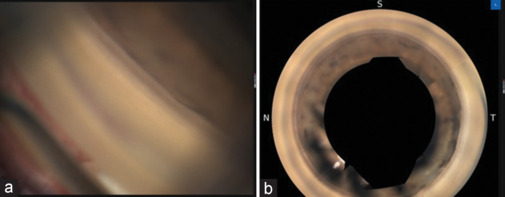
Methods: A prospective, observational study including patients who underwent PCS at Hospital Santa Maria, Lisbon, from March to October 2022. AGI was done at the preoperative visit and 6-weeks postoperative. The images were analyzed using NAVIS-EX Software (NIDEK CO., Japan) by a masked observer. Each eye was divided into four quadrants and their morphology and pigmentation was graded according to the Shaffer and modified Scheie classifications, respectively.
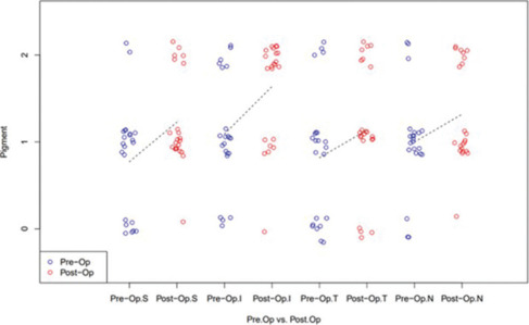
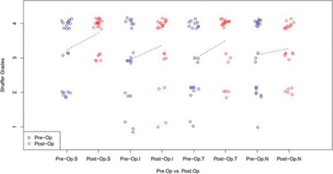
Results: Twenty-two eyes from 21 patients (62% female) were included, with a mean age of 74.5 ± 7.9 years. Baseline AGI identified 7 (33%) patients with angle closure. PCS led to all patients achieving open angle status and lower preoperative Shaffer grades achieved greater postoperative improvement. Statistically significant ICA widening was found in the superior (P = 0.004), inferior (P = 0.008), and temporal (P = 0.023) quadrants, but not the nasal quadrant (P = 0.21). Similarly, a statistically significant pigmentation increase was found in the superior (P = 0.008), inferior (P = 0.002), and temporal (P = 0.016) quadrants and less pigmented baseline quadrants showed a greater gain. The most significant pigmentation gain was in the inferior quadrant.
Conclusion: Unlike other imaging modalities, AGI with GS-1 can detect changes in ICA morphology and pigmentation after routine cataract surgery, mainly in the superior, inferior, and temporal quadrants.
期刊介绍:
Saudi Journal of Ophthalmology is an English language, peer-reviewed scholarly publication in the area of ophthalmology. Saudi Journal of Ophthalmology publishes original papers, clinical studies, reviews and case reports. Saudi Journal of Ophthalmology is the official publication of the Saudi Ophthalmological Society and is published by King Saud University in collaboration with Elsevier and is edited by an international group of eminent researchers.

 求助内容:
求助内容: 应助结果提醒方式:


